. 2018 Jan 29;9(1):420.
doi: 10.1038/s41467-017-02770-z.

Molecular basis for the specific and multivariant recognitions of RNA substrates by human hnRNP A2/B1

Affiliations

Molecular basis for the specific and multivariant recognitions of RNA substrates by human hnRNP A2/B1

Baixing Wu et al. Nat Commun. .

Abstract

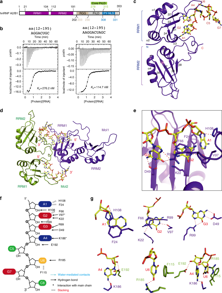
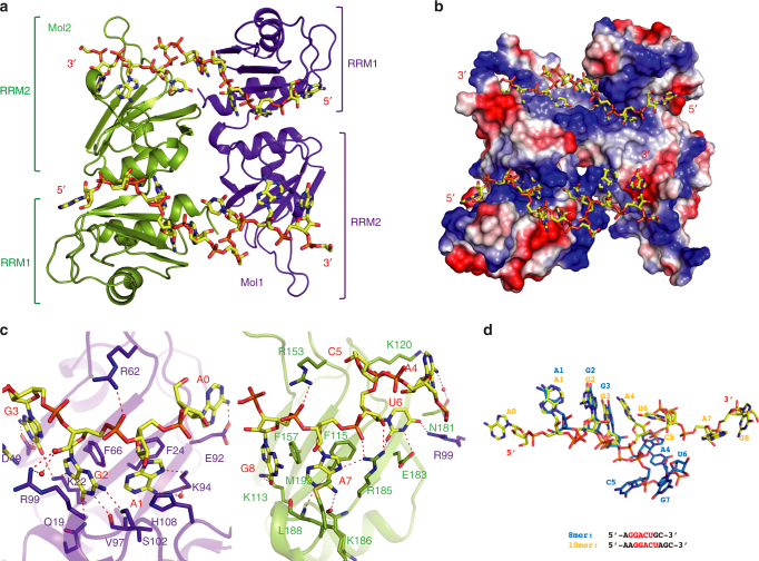
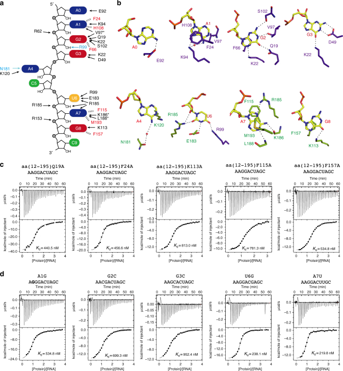
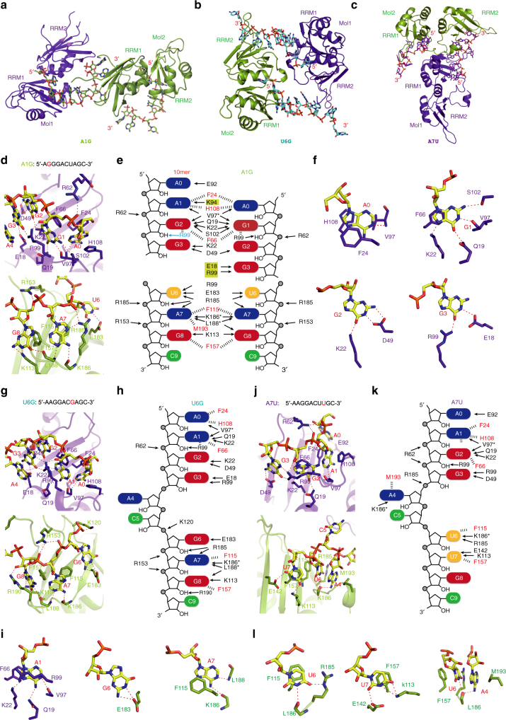
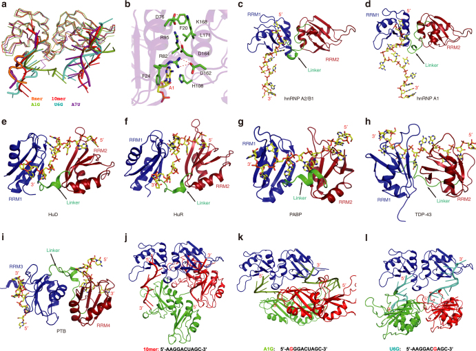
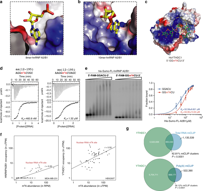
Human hnRNP A2/B1 is an RNA-binding protein that plays important roles in many biological processes, including maturation, transport, and metabolism of mRNA, and gene regulation of long noncoding RNAs. hnRNP A2/B1 was reported to control the microRNAs sorting to exosomes and promote primary microRNA processing as a potential m6A "reader." hnRNP A2/B1 contains two RNA recognition motifs that provide sequence-specific recognition of RNA substrates. Here, we determine crystal structures of tandem RRM domains of hnRNP A2/B1 in complex with various RNA substrates, elucidating specific recognitions of AGG and UAG motifs by RRM1 and RRM2 domains, respectively. Further structural and biochemical results demonstrate multivariant binding modes for sequence-diversified RNA substrates, supporting a RNA matchmaker mechanism in hnRNP A2/B1 function. Moreover, our studies in combination with bioinformatic analysis suggest that hnRNP A2/B1 may mediate effects of m6A through a "m6A switch" mechanism, instead of acting as a direct "reader" of m6A modification.


Conflict of interest statement

The authors declare no competing financial interests.

Figures

Fig. 1
Fig. 1
Overview of the structure and ITC of hnRNP A2/B1 in complex with 8mer RNA. a Schematic representation of the domain architecture of hnRNP A2/B1. RRM: RNA recognition motif, PrLD: prion-like domain, NLS: nuclear location signal, RGG: arginine-glycine-glycine box. b ITC results of hnRNP A2/B1(12–195) with 8mer and 10mer RNA targets. Solid lines indicate nonlinear least-squares fit the titration curve, with ΔH (binding enthalpy kcal mol−1), Ka (association constant), and N (number of binding sites per monomer) as variable parameters. Calculation values for Kd (dissociation constant) are indicated. c Cartoon representation of RRMs in complex with 8mer RNA. The RNA backbone is colored in yellow shown by stick. The RRMs are colored in purple-blue. d Molecules from two adjacent asymmetric units. The molecule from another asymmetric unit is colored in green. e Intermolecular contacts between RRM1 and 8mer RNA from A1 to G3. All dashed lines in this study indicate distance <3.2 Å. f Schematic showing RRMs interactions with 8mer RNA 5′-AGGACUGC-3′. g Close-up view showing the specific recognition of A1, G2, G3, A4, and U6
Fig. 2
Fig. 2
Overview of RRMs in complex with 10mer RNA. a Cartoon representation of RRMs in complex with 10mer RNA 5′-AAGGACUAGC-3′. The RNA backbone is colored in yellow shown by stick. The molecule from the adjacent asymmetric unit is colored in green. b Surface representation of RRMs–10mer complex. c Intermolecular contacts between RRM1 and residues of 10mer RNA 5′-AAGG-3′, and RRM2 with residues of RNA 5′-ACUAGC-3′. d Superposition of 8mer and 10mer RNA substrates. The 8mer RNA 5′-AGGACUGC-3′ is colored in marine and the 10mer RNA 5′-AAGGACUAGC-3′ is colored in yellow
Fig. 3
Fig. 3
Detailed interactions between RRMs and 10mer RNA. a Schematic showing RRMs interactions with 10mer RNA sequence. b Close-up view showing the specific recognition from A1 to G8. c Mutagenesis study by ITC experiments between protein mutants and 10mer RNA substrate. d ITC experiments between hnRNP A2/B1 RRMs and RNA mutants A1G, G2C, G3C, U6G, and A7U
Fig. 4
Fig. 4
RNA mutants indicate mutivariant-binding mode of hnRNP A2/B1 RRMs. a Structure of RRMs (12–195) in complex with A1G-RNA 5′-AGGGACUAGC-3′. b Structure of RRMs (12–195) in complex with U6G RNA 5′-AAGGACGAGC-3′. c Structure of RRMs (12–195) in complex with A7U-RNA 5′-AAGGACUUGC-3′. d Intermolecular contacts between RNA and RRMs in A1G complex. RRM1 is colored in purple-blue, RRM2 is colored in green. e Schematic representation of the comparison of different intermolecular interactions between 10mer RNA 5′-AAGGACUAGC-3′ and A1G-RNA 5′-AGGGACUAGC-3′. f Close-up view showing the specific recognition from A1 to G3. g Intermolecular contacts between RNA and RRMs in U6G complex. h Schematic representation of intermolecular interactions in the U6G complex. i Close-up view showing the specific recognition of A1, G6, and A7 in U6G complex. j Intermolecular contacts between RNA and RRMs in A7U complex. k Schematic representation of intermolecular interactions in A7U complex. l Close-up view showing the specific recognition of U6, U7, and the stacking interactions involved in A4, U6, F157, and M193 in A7U complex
Fig. 5
Fig. 5
The RNA binding by RRMs adopts an antiparallel mode. a Superimposition of different structure complex. 8mer-RNA: 5′-AGGACUGC-3′ is colored in orange, 10mer-RNA: 5′-AAGGACUAGC-3′ is colored in red, A1G-RNA: 5′-AGGGACUAGC-3′ is colored in green, U6G: RNA 5′-AAGGACGAGC-3′ and A7U-RNA: 5′-AAGGACUUGC-3′ are colored in cyan and purple, respectively. b Interactions between RRM1 and RRM2; the amino acids participating in interactions are colored in green and RNA is colored in yellow. c The overall structure of hnRNP A2/B1-10mer RNA complex. d hnRNP A1 in complex with DNA. eh The overall structure of HuD-RNA, HuR-RNA, PABP-RNA, and TDP-43-RNA. From c to h, RRM1 is colored in blue and RRM2 is colored in red, the linker is colored in green pointed out with a black arrow, the RNA backbone is colored in yellow shown by stick. i The overall structure of PTB–RNA complex with an antiparallel RNA-binding mode. RRM3 is colored in blue and RRM4 is colored in red, other labels are the same as from c to h. jl Crystal packing interactions in 10mer, A1G and U6G. To illustrate the detailed packing interactions of hnRNP A2/B1 carrying two antiparallel RNA stands with other hnRNP A2/B1 molecules, three hnRNP A2/B1 molecules and two RNA strands of each complex are selected to show
Fig. 6
Fig. 6
hnRNP A2/B1 does not specifically recognize m6A-modified RNA. a Surface representation of the environment around A4 in 8mer RNA complex. b Surface representation of the environment around A4 in 10mer RNA complex. c Surface representation of the canonical N6-methylated adenosine binding mode in HsYTHDC1. The aromatic cage is circled with yellow dashline. d ITC data of hnRNP A2/B1(12–195) with 8mer and 10mer RNA targets carried N6-methylated adenosine. e EMSA experiment shows the binding affinity of full-length hnRNP A2/B1 with 5′-FAM-labeled RNA substrates with or without m6A modification. Uncropped gel image is shown in Supplementary Fig. 7e. The data represent the mean of three independent experiments, with standard deviation (SD) values indicated by error bars. f YTHDC1 shows preferential binding to m6A sites in nuclear RNA compared to hnRNP A2/B1. For hnRNP A2/B1, the m6A-Seq reads that overlapped with each m6A site was plotted on the x-axis, and the HITS-CLIP reads that overlap with each site were plotted on the y-axis. A similar analysis was used to examine YTHDC1 binding at these m6A sites. miCLIP reads that overlapped with the m6A sites were plotted on the x-axis, and the YTHDC1 iCLIP reads that overlapped with the m6A sites were plotted on the y-axis. g YTHDC1-m6A tag cluster overlap. A Venn diagram indicating the cluster overlap is shown. Roughly, 43 and 56% of miCLIP tag clusters from total cellular RNA and poly(A) RNA showed a significant overlap with the YTHDC1 iCLIP clusters, respectively

References

    1. Keene JD. RNA regulons: coordination of post-transcriptional events. Nat. Rev. Genet. 2007;8:533–543. doi: 10.1038/nrg2111. - DOI - PubMed
    1. Glisovic T, Bachorik JL, Yong J, Dreyfuss G. RNA-binding proteins and post-transcriptional gene regulation. FEBS Lett. 2008;582:1977–1986. doi: 10.1016/j.febslet.2008.03.004. - DOI - PMC - PubMed
    1. He Y, Smith R. Nuclear functions of heterogeneous nuclear ribonucleoproteins A/B. Cell. Mol. Life Sci. 2009;66:1239–1256. doi: 10.1007/s00018-008-8532-1. - DOI - PMC - PubMed
    1. Gabut M, Chaudhry S, Blencowe BJ. SnapShot: the splicing regulatory machinery. Cell. 2008;133:192 e1. doi: 10.1016/j.cell.2008.03.010. - DOI - PubMed
    1. Cook KB, Kazan H, Zuberi K, Morris Q, Hughes TR. RBPDB: a database of RNA-binding specificities. Nucleic Acids Res. 2011;39:D301–D308. doi: 10.1093/nar/gkq1069. - DOI - PMC - PubMed

Publication types

MeSH terms