. 2018 Jan 29;8(1):1799.
doi: 10.1038/s41598-018-20064-2.

Preferential amplification of a human mitochondrial DNA deletion in vitro and in vivo

Affiliations

Preferential amplification of a human mitochondrial DNA deletion in vitro and in vivo

Oliver M Russell et al. Sci Rep. .

Abstract

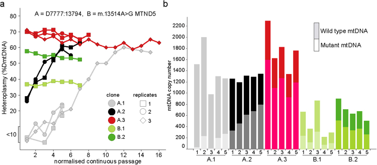
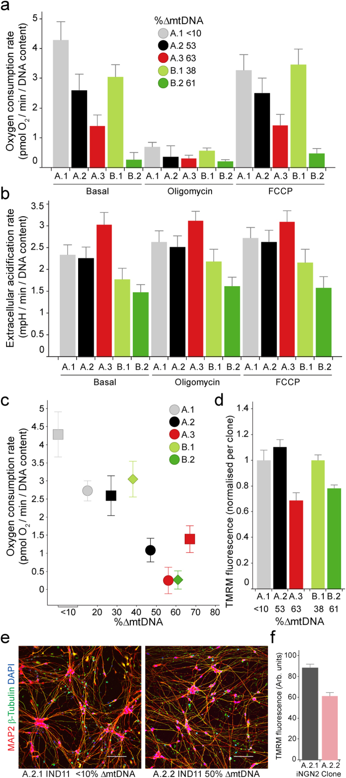
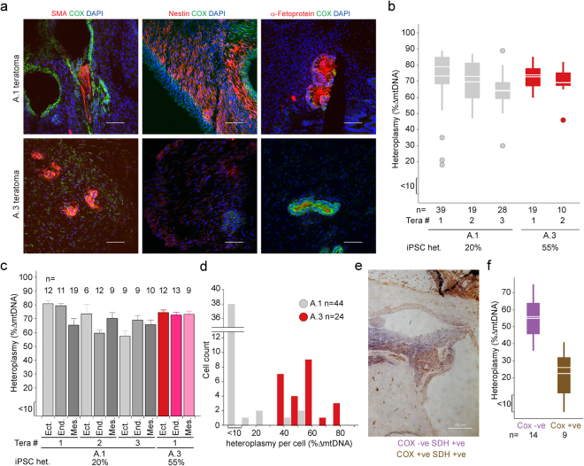
We generated induced pluripotent stem cells (iPSCs) from patient fibroblasts to yield cell lines containing varying degrees of heteroplasmy for a m.13514 A > G mtDNA point mutation (2 lines) and for a ~6 kb single, large scale mtDNA deletion (3 lines). Long term culture of the iPSCs containing a single, large-scale mtDNA deletion showed consistent increase in mtDNA deletion levels with time. Higher levels of mtDNA heteroplasmy correlated with increased respiratory deficiency. To determine what changes occurred in deletion level during differentiation, teratomas comprising all three embryonic germ layers were generated from low (20%) and intermediate heteroplasmy (55%) mtDNA deletion clones. Regardless of whether iPSCs harbouring low or intermediate mtDNA heteroplasmy were used, the final levels of heteroplasmy in all teratoma germ layers increased to a similar high level (>60%). Thus, during human stem cell division, cells not only tolerate high mtDNA deletion loads but seem to preferentially replicate deleted mtDNA genomes. This has implications for the involvement of mtDNA deletions in both disease and ageing.


Conflict of interest statement

The authors declare that they have no competing interests.

Figures

Figure 1
Figure 1
Molecular characterisation of mtDNA heteroplasmy and copy number after iPSC generation. (a) Heteroplasmy during continuous passages of iPSCs, following reprogramming of two patient fibroblasts carrying mtDNA Δ7777:13794 at 17% (A), and m.13514 A > G at 55% (B). A.1 (grey), A.2 (black), A.3 (red), B.1 (light green) and B.2 dark green) clones were continuously passaged and heteroplasmy assessed. (b) Values for wild type and mutant copy number (CN) determined from iPSCs from each replicate of the first 5 continuous passages in (a).
Figure 2
Figure 2
Heteroplasmy level correlates with mitochondrial phenotypes in iPSCs and iPSC-derived neurons. A.1 (grey), A.2 (black), A.3 (red), B.1 (light green) and B.2 dark green) iPSCs were assessed for (a) basal and maximal (FCCP) mitochondrial oxygen consumption (OCR; values corrected for non-mitochondrial OCR), and extracellular acidification (ECAR) as a proxy for glycolysis (b), and basal OCR compared to heteroplasmy (2 different passages per clone), (c) and (d) mitochondrial inner membrane potential (ψ) as visualized by TMRM staining. iNGN2 containing A.2 sub-clones A.2.1 and A.2.2 with <10% and 50% heteroplasmy respectively were differentiated for 11 (IND11) days and assessed for (e) neuronal markers using immunofluorescence (scale 10 µm) and (f) mitochondrial membrane potential using TMRM fluorescence. (a–c) mean values with s.d. n = 8 (biological replicates), (d) and (f) mean and s.e.m. (d) A.1 n = 10, A.2 n = 10, A.3 n = 10, B.1 n = 40, B.2 n = 40. (f) A.2.1 n = 93; A.2.2 n = 89.
Figure 3
Figure 3
iPSCs carrying low or intermediate levels of heteroplasmy form teratomas in which all three germlayers carry high levels of heteroplasmy. NOD/SCID γ mice were inoculated with A.1 or A.3 iPSCs (20% and 55% heteroplasmy respectively) and the resulting teratomas were assessed for (a) germlayer markers smooth muscle actin (SMA) (mesoderm), Nestin (ectoderm) and α-fetoprotein (endoderm) (scale 10 µm). (b) Heteroplasmy was determined for 5 Teratomas – 3 formed using A.1 iPSCs (grey) and 2 formed from A.3 iPSCs. (c) Germlayers were laser microdissected to assess mean and s.e.m. for heteroplasmy from 4 fully differentiated teratomas – 3 formed from A.1 (grey shades) and 1 formed from A.3 iPSC (red shades). (d) Single cell heteroplasmy analysis of A.1 and A.3 iPSCs (heteroplasmy scores binned into 15 groups) (e) Following sequential COX/SDH histochemistry, regions of teratoma demonstrating COX-reactivity (brown) or COX deficiency (purple) were laser microdissected and (f) mtDNA heteroplasmy levels determined. (b) and (f) mean – dotted line; median – solid line; wide box - 1st and 3rd quartiles; circles – outlier values.

References

    1. Yoon YG, Koob MD, Yoo YH. Re-engineering the mitochondrial genomes in mammalian cells. Anat. Cell Biol. 2010;43:97–109. doi: 10.5115/acb.2010.43.2.97. - DOI - PMC - PubMed
    1. Ma H, et al. Metabolic rescue in pluripotent cells from patients with mtDNA disease. Nature. 2015;524:234–238. doi: 10.1038/nature14546. - DOI - PubMed
    1. Fujikura J, et al. Induced pluripotent stem cells generated from diabetic patients with mitochondrial DNA A3243G mutation. Diabetologia. 2012;55:1689–1698. doi: 10.1007/s00125-012-2508-2. - DOI - PubMed
    1. Hamalainen RH, et al. Tissue- and cell-type-specific manifestations of heteroplasmic mtDNA 3243A > G mutation in human induced pluripotent stem cell-derived disease model. Proc. Natl. Acad. Sci. USA. 2013;110:E3622–30. doi: 10.1073/pnas.1311660110. - DOI - PMC - PubMed
    1. Folmes CDL, et al. Disease-causing mitochondrial heteroplasmy segregated within induced pluripotent stem cell clones derived from a MELAS patient. Stem cells (Dayton, Ohio) 2013;31:1298–1308. doi: 10.1002/stem.1389. - DOI - PMC - PubMed

Publication types

Substances

LinkOut - more resources