. 2018 Jan 29;8(1):1765.
doi: 10.1038/s41598-018-20173-y.

Immunomodulatory capacity of the serotonin receptor 5-HT2B in a subset of human dendritic cells

Affiliations

Immunomodulatory capacity of the serotonin receptor 5-HT2B in a subset of human dendritic cells

Attila Szabo et al. Sci Rep. .

Abstract

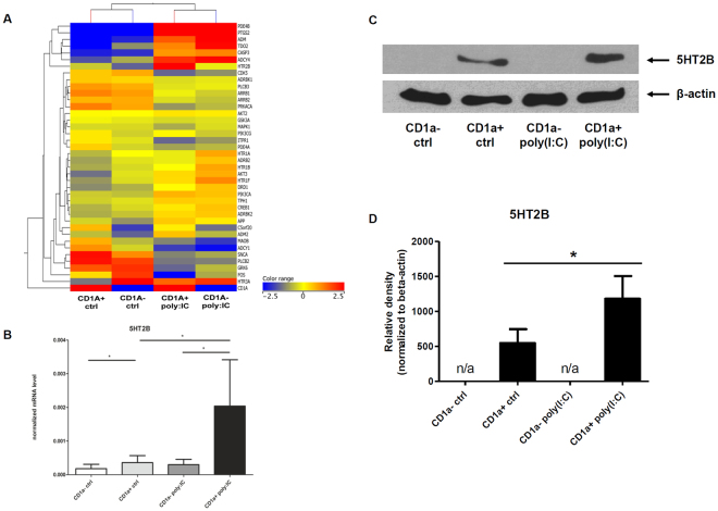
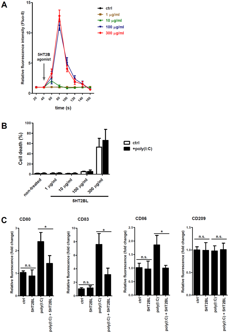
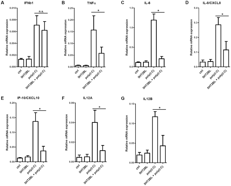
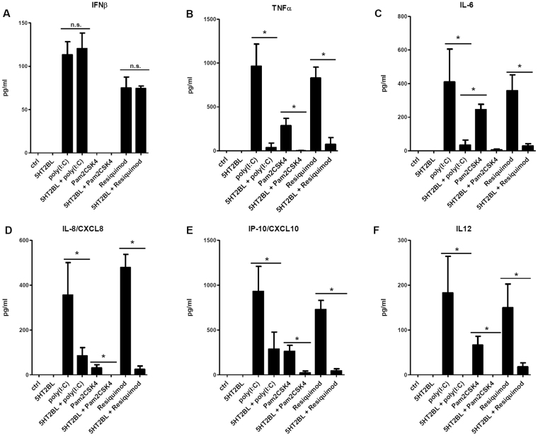
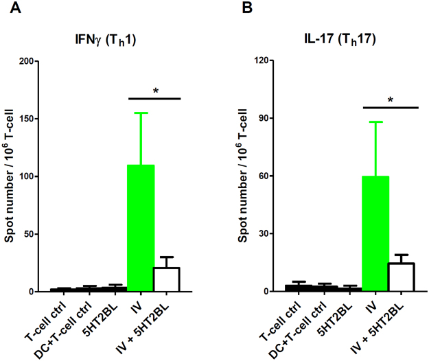
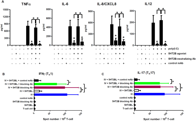
Serotonin is a monoamine neurotransmitter that signals through a wide array of receptors (5-HT1-7) many of which are also involved in immune processes. Dendritic cells (DCs) are crucial players in immune defense by bridging innate and adaptive immune responses via their vast repertoire of pattern recognition receptors and antigen-presenting capability. Although serotonin is known to influence immunity at many levels, cell type-specific expression and function of its receptors remains poorly understood. Here we aimed to study 5-HT1-7 expression and function in CD1a- and CD1a+ human monocyte-derived DCs (moDCs). We found that the 5-HT2B receptor-subtype is solely expressed by the inflammatory CD1a+ moDC subset. Specific 5-HT2B activation potently inhibited TLR2, TLR3, and TLR7/8-induced proinflammatory cytokine and chemokine (TNF-α, IL-6, IL-8, IP-10, IL-12) but not type I interferon-β responses. 5-HT2B agonism also interfered with the polarization of CD1a+ moDC-primed CD4+ T cells towards inflammatory Th1 and Th17 effector lymphocytes. Here we report the subset-specific expression and immunomodulatory function of 5-HT2B in human moDCs. Our results expand the biological role of 5-HT2B which may act not only as a neurotransmitter receptor, but also as an important modulator of both innate and adaptive immune responses.


Conflict of interest statement

The authors declare that they have no competing interests.

Figures

Figure 1
Figure 1
Distinct expression of 5-HT2B in human monocyte-derived dendritic cell subsets MoDCs were differentiated in the presence of GM-CSF and IL-4 as described in Methods. (AB) Heat map of relative mRNA expressions of serotonin receptors, transporters, metabolic enzymes and other factors involved in 5-HT signaling (panel ‘A’). Gene expressions of five independent donors were measured by TLDA array. Figure 1B represents normalized expression levels of 5-HT2B, values were extracted from the heat map on Fig. 1A. Mean ± SEM values of 5 donors are represented. (C) Western blot analysis of 5-HT2B protein expression in CD1a and CD1a+ DCs. Activated cells were treated with 20 µg/ml polyI:C for 24 h before sampling. Results of a representative experiment out of 6 is shown. (D) Densitometry data show the Mean ± SEM values of 5-HT2B protein expressions in six independent donors. Asterisk represents p values < 0.05.
Figure 2
Figure 2
Specific activation of 5-HT2B results in phenotypical and functional changes in CD1a+ DCs. CD1a+ moDCs were treated with 1–300 µg/ml of the selective 5-HT2B receptor ligand BW723C86 hydrochloride (5HT2BL, 5-HT2B agonist), and/or with 20 µg/ml polyI:C. (A) Changes in intracellular (IC) calcium levels were monitored by the fluorescence tracer Fluo-8 as in Methods. Time kinetics of IC Ca2+ alterations in non-activated control (ctrl) or in 1–300 µg/ml BW723C86 treated cells are shown. Data of triplicate measurements of four independent donors are represented as Mean ± SEM. (B) Rate of cell death following 5-HT2B activation by increasing concentrations of BW723C86. Induction of apoptosis was evaluated by Annexin V-FITC staining after 24 h. Results of four independent donors are shown as Mean ± SEM; black bars mean 20 µg/ml polyI:C co-treated cultures whereas empty bars represent non-co-treated controls. (C) Following 12 h stimulation with 20 µg/ml polyI:C and/or 100 µg/ml BW723C86 (5HT2BL) the expression levels of CD80, CD83, and CD86 cell surface proteins were analyzed by flow cytometry. Relative fluorescence intensity values were calculated using the respective isotype-matched control antibodies. The bars represent fold changes compared to the untreated control (ctrl) and data are expressed as the Mean ± SEM of four (CD80, 83, 86) or three (CD209) independent experiments. *p < 0.05; n.s. means ‘non-significant’.
Figure 3
Figure 3
Specific 5-HT2B stimulation interferes with the inflammatory response of human dendritic cells. Human CD1a+ moDCs were activated by 20 µg/ml polyI:C and/or 100 µg/ml BW723C86 (5HT2BL), and the expressions of inflammatory cytokine, chemokine, and type I interferon genes were assessed by Q-PCR. Relative mRNA expression data of triplicate measurements of six independent donors are shown as Mean ± SEM. ‘ctrl’ = non-treated control; *shows statistical significance at p < 0.05; n.s. means ‘non-significant’.
Figure 4
Figure 4
The ligation of 5HT2B alters the cytokine profile of human moDCs upon inflammatory stimulation. Human CD1a+ moDCs were activated by 20 µg/ml polyI:C (TLR3 agonist), 10 ng/ml Pam2CSK4 (TLR2 ligand), 10 µg/ml Resiquimod (TLR7/8 agonist) and/or 100 µg/ml BW723C86 (5HT2BL). Inflammatory cytokine, chemokine, and type I interferon production was detected by ELISA in culture supernatants as detailed in Methods. Results represent the Mean ± SEM of triplicates of six (TLR3 activation) or three (TLR2 and TLR7/8 activation) independent donors. *p < 0.05; n.s. means ‘non-significant’.
Figure 5
Figure 5
Activation of the 5-HT2B receptor of human moDCs inhibits inflammatory adaptive immune responses. Human CD1a+ moDCs were activated by inactivated influenza virus (IV) for 24 h, washed, and then co-cultured with naive autologous CD4+ T lymphocytes for 4 days. The number of primed, IFNγ or IL-17 secreting T cells was assessed by ELISPOT assay. T cells alone (T-cell ctrl) or cultured with immature DCs (DC + T-cell ctrl) were used as controls. Induction of IFNγ (A) or IL-17 (B) production of autologous naive CD4+ T cells primed by CD1a+ moDC loaded with inactivated influenza virus (IV) is shown. 100 µg/ml BW723C86 (5HT2BL) was added to the moDCs alone for 24 h (black bars), or in combination with influenza virus (IV + 5HT2BL; empty bars). Green bars represent influenza virus-only activation (IV). Data represent Mean + SEM values of triplicate measurements of six independent donors. Asterisk indicates statistical significance (p < 0.05).
Figure 6
Figure 6
5-HT2B receptor-neutralization abrogates the inhibitory effect of BW723C86 on the cytokine profile and Th1/Th17 cell-priming capacity of activated moDCs. Cells were co-cultured, activated, and the levels of secreted cytokines as well as the number of IFNγ or IL-17 secreting T cells was assessed by ELISA and ELISPOT as in Figs 4 and 5. (A) Non-treated, 100 µg/ml BW723C86, and 5-HT2B-neutralizing antibody-treated cells were used as negative controls. Cells treated with 20 µg/ml polyI:C served as positive controls. Co-treatments with polyI:C + BW723C86 were done as in Figs 3 and 4. 5-HT2B receptor-neutralizing or isotype-matched monoclonal antibodies (control mAb) were always added to the cultures 30 minutes prior to activation. Supernatants of cultures were collected after 12 h and were measured by ELISA. Concentration of the secreted cytokines and chemokines are shown as Mean ± SEM values of three independent donors. (BC) Blue bars represent influenza antigen-loaded DCs co-cultured with autologous naive CD4+ T cells (positive control). White bars show moDC co-activation with IV + 5HT2BL and subsequent T cell co-culturing. Claret bars (IV + 5HT2B blocking Ab) and green bars (IV + 5HT2BL + 5HT2B blocking antibody) demonstrate cultures that had been pre-treated with 5-HT2B blocking antibody for 30 minutes prior to virus-loading (claret) or virus + BW723C86 co-treatment (green), respectively. DCs treated only with either 5HT2BL, the receptor-specific blocking antibody or an isotype-matched monoclonal antibody (control mAb), and co-cultured with CD4+ T cells were used as controls. Data represent Mean + SEM values of triplicate measurements of three independent donors. Asterisk shows statistical significance (p < 0.05).

References

    1. Belz GT, Nutt SL. Transcriptional programming of the dendritic cell network. Nature reviews. Immunology. 2012;12:101–113. doi: 10.1038/nri3149. - DOI - PubMed
    1. Szabo A, Rajnavolgyi E. Collaboration of Toll-like and RIG-I-like receptors in human dendritic cells: tRIGgering antiviral innate immune responses. American journal of clinical and experimental immunology. 2013;2:195–207. - PMC - PubMed
    1. Worbs T, Hammerschmidt SI, Forster R. Dendritic cell migration in health and disease. Nature reviews. Immunology. 2017;17:30–48. doi: 10.1038/nri.2016.116. - DOI - PubMed
    1. Berger M, Gray JA, Roth BL. The expanded biology of serotonin. Annual review of medicine. 2009;60:355–366. doi: 10.1146/annurev.med.60.042307.110802. - DOI - PMC - PubMed
    1. Hannon J, Hoyer D. Molecular biology of 5-HT receptors. Behavioural brain research. 2008;195:198–213. doi: 10.1016/j.bbr.2008.03.020. - DOI - PubMed

Publication types

MeSH terms