. 2018 Jan 29;8(1):1809.
doi: 10.1038/s41598-018-19802-3.

Downregulation of glutamic acid decarboxylase in Drosophila TDP-43-null brains provokes paralysis by affecting the organization of the neuromuscular synapses

Affiliations

Downregulation of glutamic acid decarboxylase in Drosophila TDP-43-null brains provokes paralysis by affecting the organization of the neuromuscular synapses

Giulia Romano et al. Sci Rep. .

Abstract

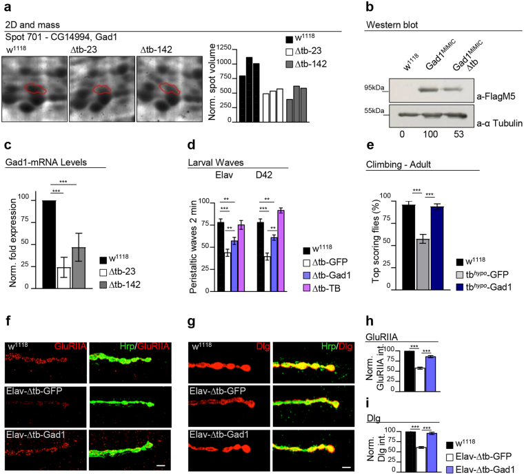
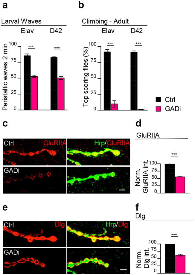
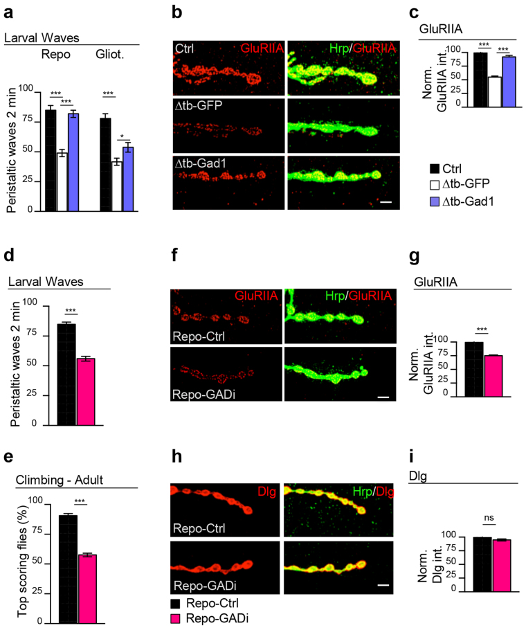
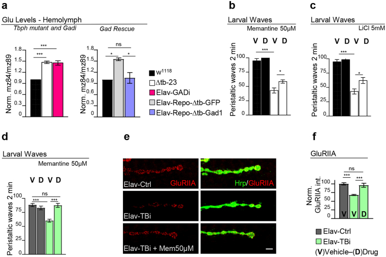
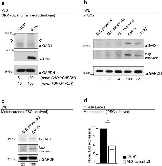
Amyotrophic lateral sclerosis is a progressive neurodegenerative disease that affects the motor system, comprised of motoneurons and associated glia. Accordingly, neuronal or glial defects in TDP-43 function provoke paralysis due to the degeneration of the neuromuscular synapses in Drosophila. To identify the responsible molecules and mechanisms, we performed a genome wide proteomic analysis to determine differences in protein expression between wild-type and TDP-43-minus fly heads. The data established that mutant insects presented reduced levels of the enzyme glutamic acid decarboxylase (Gad1) and increased concentrations of extracellular glutamate. Genetic rescue of Gad1 activity in neurons or glia was sufficient to recuperate flies locomotion, synaptic organization and glutamate levels. Analogous recovery was obtained by treating TDP-43-null flies with glutamate receptor antagonists demonstrating that Gad1 promotes synapses formation and prevents excitotoxicity. Similar suppression of TDP-43 provoked the downregulation of GAD67, the Gad1 homolog protein in human neuroblastoma cell lines and analogous modifications were observed in iPSC-derived motoneurons from patients carrying mutations in TDP-43, uncovering conserved pathological mechanisms behind the disease.


Conflict of interest statement

The authors declare that they have no competing interests.

Figures

Figure 1
Figure 1
Gad1 is downregulated in TBPH-mutant flies and required for synaptogenesis. (a) Two-dimensional electrophoresis analysis of adult heads in Drosophila revealed that the spot 701, corresponding to the Gad1-CG14994 protein was reduced in Δtb-23 and Δtb-142 (tbphΔ23/Δ23 and tbphΔ142/Δ142) null alleles compared to w1118controls. The bar graph indicates quantified spot intensities (volumes), after gel normalization. n = 3. (b) Western blot analysis of w1118, Gad1MiMIC (w; +/+;Mi{MIC}Gad1MI09277/+) and Gad1MiMIC − Δtb (w;tbphΔ23/tbphΔ23;Mi{MIC}Gad1MI09277/+). Third instar larval brains were probed with anti-FlagM5 and alpha-tubulin antibodies. The same membrane was probe with the two antibodies and the bands of interest were cropped. Quantification of normalized amounts was reported below each lane. n = 3. (c) Real time PCR of Gad1 transcript levels normalized on Rpl11 (housekeeping) in third instar larval brains of w1118, Δtb-23 (tbphΔ23/Δ23) and Δtb-142 (tbphΔ142/Δ142). n = 3. (d) Number of peristaltic waves between w1118, Δtb-GFP (left: elav-GAL4,tbphΔ23/tbphΔ23;UAS-GFP/ +; right: tbphΔ23/tbphΔ23;D42-GAL4/UAS-GFP), Δtb-Gad1 (left side: elav-GAL4,tbphΔ23/tbphΔ23;UAS-Gad1/+; right side: tbphΔ23/tbphΔ23;D42-GAL4/UAS-Gad1) and Δtb-TB (left side: elav-GAL4,tbphΔ23/tbphΔ23,UAS-TBPH; right side: tbphΔ23/tbphΔ23,UAS-TBPH;D42-GAL4/+). n = 25. (e) Climbing assay on 4 days old adult flies compare w1118, tbhypo-GFP (elav-GAL4,tbphΔ23/UAS-GFP;UAS-Dcr-2,TBPH-RNAi/+) and tbhypo-Gad1 (elav-GAL4,tbphΔ23/ + ;UAS-Dcr-2,TBPH-RNAi/UAS-Gad1). n = 100. (f) Confocal images of third instar NMJ terminals, muscle 6/7 second segment stained with anti-HRP (in green) and anti-GluRIIA (in red) in w1118, Elav-Δtb-GFP and Elav-Δtb-Gad1. (g) Confocal images of third instar NMJ terminals in muscle 6/7 second segment stained with anti-HRP (in green) and anti-Dlg (in red) in w1118, Elav-Δtb-GFP and Elav-Δtb-Gad1. (h-i) Quantification of GluRIIA intensity and Dlg intensity, n > 200 boutons. **p < 0.01, ***p < 0.001 calculated by one-way ANOVA. Error bars SEM. Scale bar 5 µm.
Figure 2
Figure 2
The reduction of Gad1 in neurons triggers postsynaptic assembly defects. (a) Number of peristaltic waves of Ctrl (left side: UAS-LacZ/elav-GAL4 and right side:UAS-Dcr-2/+;UAS-LacZ/+;D42-GAL4/+) and GADi (left side: elav-GAL4/+;GAD-RNAi/+ and right side: UAS-Dcr-2/+;;D42-GAL4/GAD-RNAi) larvae. n = 20. (b) Climbing assay on adult flies at day 4 of Ctrl and GADi on the left using elav-GAL4 and on the right using D42-GAL4. n = 100. (c) Confocal images of third instar NMJ terminals in muscle 6/7 second segment stained with anti-HRP (in green) and anti-GluRIIA (in red) in Ctrl and GADi (using elav-GAL4). (d) Quantification of GluRIIA intensity in Ctrl and GADi. n = 200 boutons. (e) Confocal images of third instar NMJ terminals in muscle 6/7 second segment stained with anti-HRP (in green) and anti-Dlg (in red) in Ctrl and GADi (using elav-GAL4). (f) Quantification of Dlg intensity in Ctrl and GADi. n = 200 boutons. ***p < 0.001, calculated by T-test. Error bars SEM. Scale bar 5 µm.
Figure 3
Figure 3
The glial activity of Gad1 is sufficient to ensure the postsynaptic clustering of the GluRIIA. (a) Number of peristaltic waves of Ctrl (left side: tbphΔ23/+; repo-GAL4,UAS-GFP/+; right side: tbphΔ23,gliotactin-GAL4/+; UAS-GFP/+), Δtb-GFP (left side: tbphΔ23/tbphΔ23;repo-GAL4/UAS-GFP; right side: tbphΔ23,gliotactin-GAL4/tbphΔ23;UAS-GFP/+) and Δtb-Gad1 (left side: tbphΔ23/tbphΔ23;repo-GAL4/UAS-Gad1; left side: tbphΔ23, gliotactin-GAL4/ tbphΔ23;UAS-GFP/+). n = 25. (b) Confocal images of third instar NMJ terminals in muscle 6/7 second segment stained with anti-HRP (in green) and anti-GluRIIA (in red) in Ctrl, Δtb-GFP and Δtb-Gad1 (using repo-GAL4). (c) Quantification of GluRIIA intensity. n > 200 boutons. (d) Number of peristaltic waves of Repo-Ctrl (UAS-Dcr-2/+;UAS-LacZ/ + ;repo-GAL4/+) and Repo-GADi (UAS-Dcr-2/+; +/+; repo-GAL4/GAD-RNAi) larvae. n = 20. (e) Climbing assay on adult flies at day 4 of Repo-Ctrl and Repo-GADi. n = 100. (f) Confocal images of third instar NMJ terminals in muscle 6/7 second segment stained with anti-HRP (in green) and anti-GluRIIA (in red) in Repo-Ctrl and Repo-GADi. (g) Quantification of GluRIIA intensity. n > 200 boutons. (h) Confocal images of third instar NMJ terminals in muscle 6/7 second segment stained with anti-HRP (in green) and anti-Dlg (in red) in Repo-Ctrl and Repo-GADi. (i) Quantification of Dlg intensity. n > 200 boutons. ns = not significant *p < 0.05, ***p < 0.001 calculated by one-way ANOVA in (a and c) and T-test in (d,e,g and i). Error bars SEM. Scale bar 5 µm.
Figure 4
Figure 4
Gad1 downregulation provokes glutamate excitotoxicity in TBPH mutant flies. (a) Glutamate relative concentration in the hemolymph of third instar larvae: on the left graph in w1118, ∆tb-23 (tbphΔ23/Δ23) and Elav-GADi (elav-GAL4/+;GAD-RNAi/+) and on the right graph the glutamate levels in w1118, Elav-Repo-ΔtbGFP (elav-GAL4,tbphΔ23/tbphΔ23;repo-GAL4,UAS-GFP/+) and Elav-Repo-Δtb-Gad1 (elav-GAL4,tbphΔ23/tbphΔ23;repo-GAL4,UAS-GFP/UAS-Gad1). Left graph n = 3, right graph n = 2. The axis of ordinates showed the ratio between the chosen fragment of the fragmentation spectra of glutamic acid and isotopic labelled glutamic acid. (b) Number of peristaltic waves of L3 larvae fed in food containing 50 µM Memantine “D-Drug” or containing the vehicle only “V-Vehicle”, in w1118 and ∆tb-23 (tbphΔ23/Δ23). n = 20. (c) Number of peristaltic waves of L3 larvae fed in food containing 5 mM Lithium chloride (LiCl) “D-Drug” or containing the vehicle only “V-Vehicle”, in w1118 and ∆tb-23 (tbphΔ23/Δ23). n = 20. (d) Number of peristaltic waves of L3 larvae fed in food containing 50 µM Memantine “D-Drug” or containing the vehicle only “V-Vehicle”, in Elav-Ctrl (UAS-Dcr-2/+;elav-GAL4,tbphΔ23/UAS-LacZ) and Elav-TBi (UAS-Dcr-2/+;elav-GAL4,tbphΔ23/+;TBPH-RNAi/+). n = 20. (e) Confocal images of third instar NMJ terminals in muscle 6/7 second segment stained with anti-HRP (in green) and anti-GluRIIA (in red) in Elav-Ctrl and Elav-TBi with and without the treatment. (f) Quantification of GluRIIA intensity. n > 200 boutons. ns = not significant *p < 0.05, ***p < 0.001 calculated by one-way ANOVA. Error bars SEM. Scale bar 5 µm.
Figure 5
Figure 5
The dysregulation of Gad1 is conserved in human cell lines and iPSCs ALS patients derived. (a) Western blot analysis on human neuroblastoma (SK-N-BE) cell line probed for anti-GAD1, anti-GAPDH and anti-TDP in siTDP (TDP silenced) and siLuc (Luciferase ctrl). The same membrane was probe with the three antibodies and the bands of interest were cropped. Quantification of normalized protein amount was reported below each lane, n = 3. (b) Western blot analysis on human iPSCs probed for anti-GAD1 and anti-GAPDH in three clones derived from three different ALS patients (ALS patient #1 carrying the G287S mutation; ALS patient #2 carrying the G294V mutation; ALS patient #3 carrying the G378S mutation) and in two clones derived from two different healthy subjects (Ctrl #1 Ctrl #2). The same membrane was probe with the two antibodies and the bands of interest were cropped. In the two upper panels were reported two different exposition of anti-GAD1 signal. Quantification of normalized protein amount was reported below each lane. n = 3. (c) Western blot analysis probed for anti-GAD1 and anti-GAPDH on human differentiated motoneurons derived from iPSCs of an ALS patient (ALS patient #3 carrying the G378S mutation) and a healthy control (Ctrl #1 clone ND41864). The same membrane was probe with the three antibodies and the bands of interest were cropped. In the two upper panels were reported two different exposition of anti-GAD1 signal. Quantification of normalized protein amount was reported below each lane. (d) Real time PCR of Gad1 transcript levels normalized on Gapdh (housekeeping) in human differentiated motoneurons derived from iPSCs of an ALS patient (ALS patient #3 carrying the G378S mutation) and a healthy control (Ctrl #1 clone ND41864). n = 2, **p < 0.01 calculated by T-test. Error bars SEM.

Similar articles

Cited by

References

    1. Neumann M, et al. Ubiquitinated TDP-43 in frontotemporal lobar degeneration and amyotrophic lateral sclerosis. Science. 2006;314:130–133. doi: 10.1126/science.1134108. - DOI - PubMed
    1. Arai T, et al. TDP-43 is a component of ubiquitin-positive tau-negative inclusions in frontotemporal lobar degeneration and amyotrophic lateral sclerosis. Biochem. Biophys. Res. Commun. 2006;351:602–611. doi: 10.1016/j.bbrc.2006.10.093. - DOI - PubMed
    1. Geser F, Martinez-Lage M, Kwong LK, Lee VM-Y, Trojanowski JQ. Amyotrophic lateral sclerosis, frontotemporal dementia and beyond: the TDP-43 diseases. J. Neurol. 2009;256:1205–1214. doi: 10.1007/s00415-009-5069-7. - DOI - PMC - PubMed
    1. Wu L-S, et al. TDP-43, a neuro-pathosignature factor, is essential for early mouse embryogenesis. Genes. N. Y. N 2000. 2010;48:56–62. - PubMed
    1. Sephton CF, et al. TDP-43 is a developmentally regulated protein essential for early embryonic development. J. Biol. Chem. 2010;285:6826–6834. doi: 10.1074/jbc.M109.061846. - DOI - PMC - PubMed

Publication types

MeSH terms