. 2017 Oct-Dec;27(4):488-495.
doi: 10.4103/ijri.IJRI_204_16.

Endovascular uterine artery interventions

Affiliations

Endovascular uterine artery interventions

Chandan J Das et al. Indian J Radiol Imaging. 2017 Oct-Dec.

Abstract

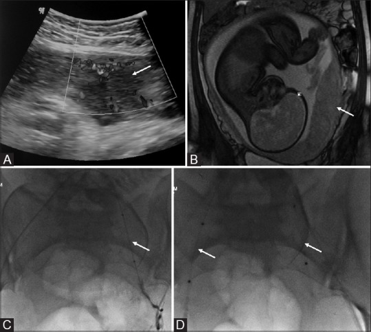
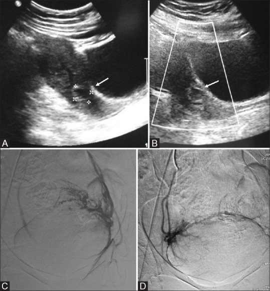
Percutaneous vascular embolization plays an important role in the management of various gynecologic and obstetric abnormalities. Transcatheter embolization is a minimally invasive alternative procedure to surgery with reduced morbidity and mortality, and preserves the patient's future fertility potential. The clinical indications for transcatheter embolization are much broader and include many benign gynecologic conditions, such as fibroid, adenomyosis, and arteriovenous malformations (AVMs), as well as intractable bleeding due to inoperable advanced-stage malignancies. The most well-known and well-studied indication is uterine fibroid embolization. Uterine artery embolization (UAE) may be performed to prevent or treat bleeding associated with various obstetric conditions, including postpartum hemorrhage (PPH), placental implantation abnormality, and ectopic pregnancy. Embolization of the uterine artery or the internal iliac artery also may be performed to control pelvic bleeding due to coagulopathy or iatrogenic injury. This article discusses these gynecologic and obstetric indications for transcatheter embolization and reviews procedural techniques and outcomes.

Keywords: Angiography; embolization; uterine artery.


Conflict of interest statement

There are no conflicts of interest.

Figures

Figure 1(A-C)
Figure 1(A-C)
Schematic image showing hooking of left internal iliac artery in a conventional way (A), followed by creation of a “Waltman loop” (B) by rotating the catheter within the aorta and then pulling it towards ipsilateral side of puncture to catheterize the ipsilateral internal iliac artery (C)
Figure 2(A-H)
Figure 2(A-H)
Axial (A) and sagittal (B) T2-weighted magnetic resonance imaging (MRI) images of a premenopausal patient with pelvic pain and menorrhagia reveal multiple leiomyomas with heterogeneous signal causing indentation of the endometrium (arrow in A and B). (C and D) digital subtraction angiography (DSA) images obtained with selective injections of the right (C) and left (D) uterine arteries demonstrate an enlarged, hypervascular uterus with multiple tortuous branches and lesion blush bilaterally. Bilateral uterine artery embolization (UAE) was performed by using polyvinyl alcohol (PVA) particles with a diameter of 500–700 μm. (E and F) Post-UAE DSA images show occlusion of multiple tortuous branches of both the right (E) and left (F) uterine arteries with static column of contrast in uterine arteries and almost complete disappearance of the lesion blush. Contrast enhanced MRI (G and H) done after 1 month of embolization showing complete nonenhancement of the fibroid (black arrow) surrounded by normally enhancing uterine wall (asterisk)
Figure 3(A-C)
Figure 3(A-C)
(A) Sagittal T2W magnetic resonance imaging (MRI) image of a 28-year-old woman shows a large intramural heterogeneous signal intensity uterine mass with prominent flow voids suggestive of an arteriovenous malformation (AVM). (B) Left internal iliac DSA showed high-flow intrauterine AVM. Embolization was performed with a combination of PVA particles and glue to occlude the vessels at the nidus. (C) Postembolization internal iliac digital subtraction angiography (DSA) image (same patient as in b) shows no filling of the AVM
Figure 4(A-F)
Figure 4(A-F)
(A) Transvaginal ultrasound image of a 50-year-old woman presenting with menorrhagia reveals an enlarged uterus with heterogeneous echopattern. (B) Sagittal T2W magnetic resonance imaging (MRI) shows extensive adenomyosis with diffuse low T2 SI thickening of the junctional zone (arrow). (C and D) digital subtraction angiography (DSA) images obtained with selective injections of the right (C) and left (D) uterine arteries demonstrate an enlarged, hypervascular uterus. Bilateral uterine artery embolization (UAE) was performed by using polyvinyl alcohol (PVA) particles with a diameter of 500–700 μm. (E, F) Post-UAE DSA images show occlusion of multiple tortuous branches of both the right (E) and the left (F) uterine arteries
Figure 5(A-D)
Figure 5(A-D)
(A) Sagittal transabdominal ultrasound (US) image shows a gap in the myometrial blood flow (arrow). (B) Coronal True fast imaging with steady state precession (TruFISP) MR image shows discontinuity of the hypointense inner myometrial layer with the placenta bulging into the myometrium (arrow) suggestive of placenta increta. (C) digital subtraction angiography (DSA) image of the same patient-showing placement of balloon within the left internal iliac artery (arrow). (D) Final fluoroscopic image demonstrates the deflated balloons in position in bilateral internal iliac arteries (arrows). The balloons were inflated immediately prior to delivery
Figure 6(A-D)
Figure 6(A-D)
(A) Transabdominal ultrasound image of a 28-year-old patient with 8 weeks amenorrhoea showing small gestational sac in the cervical canal (arrow). (B) Color Doppler reveals a ring of peripheral vascularity (arrow) suggestive of ectopic cervical pregnancy. Pre-curettage angioembolization (C and D) were performed to reduce vascularity

References

    1. Andrews RT, Spies JB, Sacks D, Worthington-Kirsch RL, Niedzwiecki GA, Marx MV, et al. Patient care and uterine artery embolization for leiomyomata. J Vasc Interv Radiol. 2009;20:S307–11. - PubMed
    1. Bulman JC, Ascher SM, Spies JB. Current concepts in uterine fibroid embolization. Radiographics. 2012;32:1735–50. - PubMed
    1. Resnick NJ, Kim E, Patel RS, Lookstein RA, Nowakowski FS, Fischman AM. Uterine artery embolization using a transradial approach: Initial experience and technique. J Vasc Interv Radiol. 2014;25:443–7. - PubMed
    1. Pelage JP, Le Dref O, Soyer P, Jacob D, Kardache M, Dahan H, et al. Arterial anatomy of the female genital tract: Variations and relevance to transcatheter embolization of the uterus. AJR Am J Roentgenol. 1999;172:989–94. - PubMed
    1. Binkert CA, Hirzel FC, Gutzeit A, Zollikofer CL, Hess T. Superior hypogastric nerve block to reduce pain after uterine artery embolization: Advanced technique and comparison to epidural anesthesia. Cardiovasc Intervent Radiol. 2015;38:1157–61. - PubMed