Review
. 2018 Jan 5:8:361.
doi: 10.3389/fendo.2017.00361. eCollection 2017.

Mechanistic Insights into Regulation of JAK2 Tyrosine Kinase

Affiliations
Review

Mechanistic Insights into Regulation of JAK2 Tyrosine Kinase

Stevan R Hubbard. Front Endocrinol (Lausanne). .

Abstract

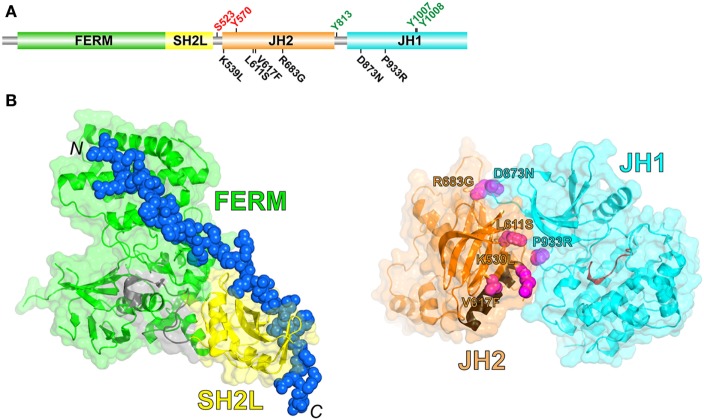
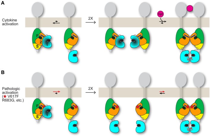
JAK2 is a member of the Janus kinase (JAKs) family of non-receptor protein tyrosine kinases, which includes JAK1-3 and TYK2. JAKs serve as the cytoplasmic signaling components of cytokine receptors and are activated through cytokine-mediated trans-phosphorylation, which leads to receptor phosphorylation and recruitment and phosphorylation of signal transducer and activator of transcription (STAT) proteins. JAKs are unique among tyrosine kinases in that they possess a pseudokinase domain, which is just upstream of the C-terminal tyrosine kinase domain. A wealth of biochemical and clinical data have established that the pseudokinase domain of JAKs is crucial for maintaining a low basal (absence of cytokine) level of tyrosine kinase activity. In particular, gain-of-function mutations in the JAK genes, most frequently, V617F in the pseudokinase domain of JAK2, have been mapped in patients with blood disorders, including myeloproliferative neoplasms and leukemias. Recent structural and biochemical studies have begun to decipher the molecular mechanisms that maintain the basal, low-activity state of JAKs and that, via mutation, lead to constitutive activity and disease. This review will examine these mechanisms and describe how this knowledge could potentially inform drug development efforts aimed at obtaining a mutant (V617F)-selective inhibitor of JAK2.

Keywords: Janus kinases; autoinhibition; cell signaling and regulation; cytokine receptor; protein tyrosine kinases.


Figures

Figure 1
Figure 1
(A) Schematic diagram of the domain organization in JAK2, shown to linear scale (1132 residues in human JAK2). Positive- and negative-regulatory phosphorylation sites (colored green and red, respectively) whose mechanisms are understood are shown above the domains. Select activating mutations in JH2 and JH1 are shown below the domains. (B) (Left) Crystal structure of the JAK1 FERM and SH2L domains in complex with a peptide representing the interferon-λ1 receptor (6) (PDB code 5L04). The FERM domain is colored green, the SH2L domain is colored yellow, the SH2L-JH2 linker is colored gray, and the receptor peptide (shown in sphere representation; the N- and C-termini are labeled) is colored blue. (Right) Crystal structure of JH2 and JH1 of TYK2 (8) (PDB code 4OLI). JH2 is colored orange, with the C helix colored brown, and JH1 is colored cyan, with the catalytic loop colored red. The positions of select activating mutations in JAK2, corresponding to those in (A), are shown in sphere representation and colored magenta (TYK2 residues shown, labeled according to the JAK2 mutations).
Figure 2
Figure 2
Possible models for JAK2 activation by cytokine and by activating mutation. JAK2 is associated with the cytoplasmic region of a cytokine receptor (gray). The four domains of JAK2 are labeled “F” for FERM, “S” for SH2L, “2” for JH2, and “1” for JH1. The integrated FERM and SH2L domains bind to the cytokine receptor. How JH2–JH1 interacts with FERM-SH2L is not known. The C helix of JH2 is represented by a dark blue rectangle, and ATP is represented by a black ellipse. (A) (Left) An equilibrium (black arrows) exists between the autoinhibited state, in which JH2 exerts an autoinhibitory interaction on JH1 (darkened), and a state in which JH1 is disengaged from JH2 and is phosphorylation-competent, with the autoinhibitory state favored for wild-type JAK2. (Middle) In the absence of cytokine, trans-phosphorylation of autoinhibited JH1 is limited. (Right) Binding of a cytokine (magenta) to the extracellular region of the receptor induces receptor dimerization, which promotes trans-phosphorylation of JH1 (red arrows). The dimerization process might also include formation of a JAK2 dimer, shown here as a JH2 dimer. (B) (Left) Activating point mutations (shown as a red star) such as V617F and R683G in JH2 shift the equilibrium from the autoinhibitory state to the phosphorylation-competent (and JAK2 dimerization-competent) state. (Middle and right) Mutant JAK2 is capable of dimerizing in the absence of cytokine. Overlap is predicted between the interfaces used by JH2 for JH1 autoinhibition and for JH2-mediated JAK2 dimerization. The activating mutation may directly disrupt the autoinhibitory interaction (e.g., R683G) or hyperstabilize the JAK2 dimer, or both (possibly V617F). Mutations that destabilize the domain structure of JH2, such as those in the ATP binding pocket (31), would suppress activating mutations (V617F, etc.) by destabilizing JAK2 dimer formation, which is necessary for activation in the absence of cytokine. Whether the putative JH2-mediated JAK2 dimer required for mutational activation in the basal state (B) is the same JAK2 dimer that may form when cytokine dimerizes the receptors (A) is not known. If homodimeric receptors such as EpoR are pre-dimerized in the basal state, then cytokine binding would rearrange the dimer (instead of dimerizing monomeric receptor-JAK2 pairs) to juxtapose the two kinase domains for trans-phosphorylation (A), possibly facilitated by a JH2-mediated interaction. An activating mutation would promote the receptor-JAK2 rearrangement through JH2-mediated JAK2 dimerization in the absence of cytokine binding (B).

References

    1. O’Shea JJ, Holland SM, Staudt LM. JAKs and STATs in immunity, immunodeficiency, and cancer. N Engl J Med (2013) 368:161–70.10.1056/NEJMra1202117 - DOI - PMC - PubMed
    1. Wang X, Lupardus P, Laporte SL, Garcia KC. Structural biology of shared cytokine receptors. Annu Rev Immunol (2009) 27:29–60.10.1146/annurev.immunol.24.021605.090616 - DOI - PMC - PubMed
    1. Levy DE, Darnell JE, Jr. Stats: transcriptional control and biological impact. Nat Rev Mol Cell Biol (2002) 3:651–62.10.1038/nrm909 - DOI - PubMed
    1. Wallweber HJ, Tam C, Franke Y, Starovasnik MA, Lupardus PJ. Structural basis of recognition of interferon-alpha receptor by tyrosine kinase 2. Nat Struct Mol Biol (2014) 21:443–8.10.1038/nsmb.2807 - DOI - PMC - PubMed
    1. McNally R, Toms AV, Eck MJ. Crystal structure of the FERM-SH2 module of human Jak2. PLoS One (2016) 11:e0156218.10.1371/journal.pone.0156218 - DOI - PMC - PubMed