. 2018 Jan 5:8:1835.
doi: 10.3389/fimmu.2017.01835. eCollection 2017.

Effects of Anti-IL-17 on Inflammation, Remodeling, and Oxidative Stress in an Experimental Model of Asthma Exacerbated by LPS

Affiliations

Effects of Anti-IL-17 on Inflammation, Remodeling, and Oxidative Stress in an Experimental Model of Asthma Exacerbated by LPS

Leandro do Nascimento Camargo et al. Front Immunol. .

Abstract

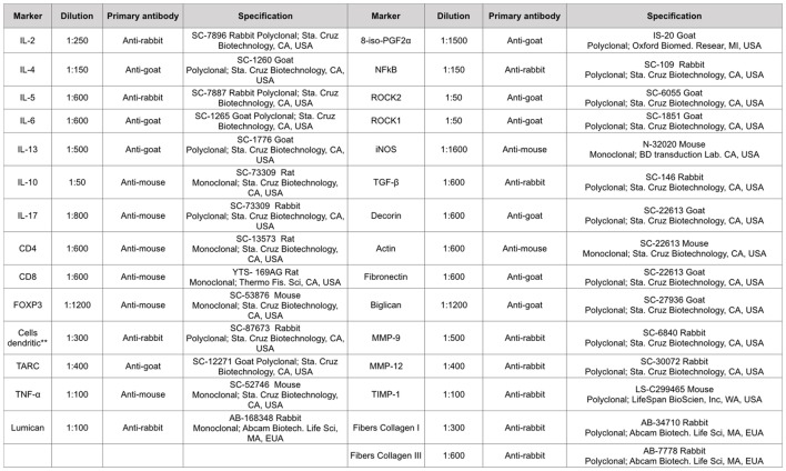
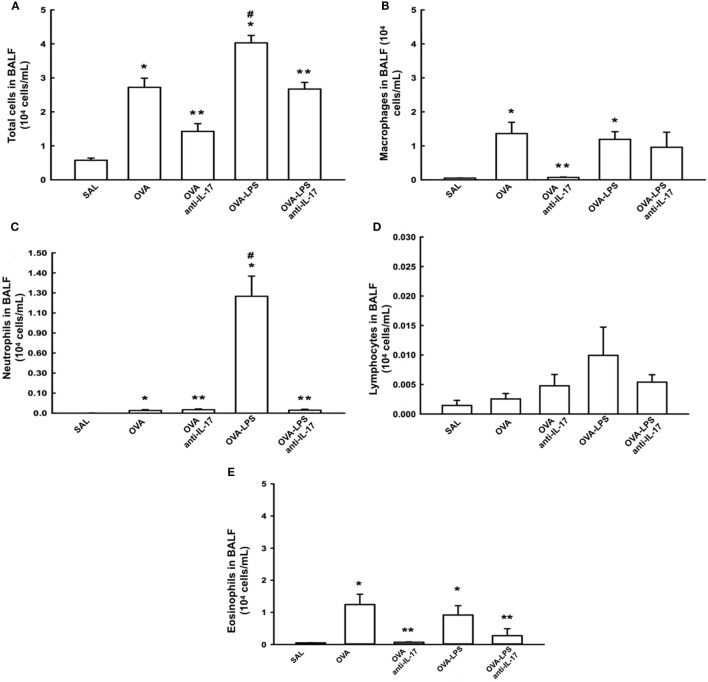
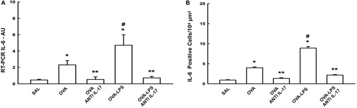
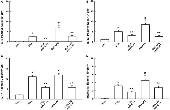
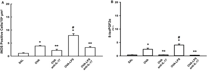
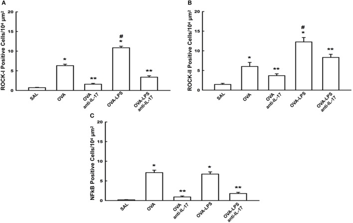
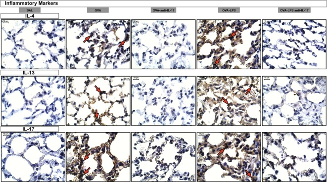
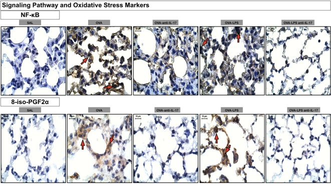
Inflammation plays a central role in the development of asthma, which is considered an allergic disease with a classic Th2 inflammatory profile. However, cytokine IL-17 has been examined to better understand the pathophysiology of this disease. Severe asthmatic patients experience frequent exacerbations, leading to infection, and subsequently show altered levels of inflammation that are unlikely to be due to the Th2 immune response alone. This study estimates the effects of anti-IL-17 therapy in the pulmonary parenchyma in a murine asthma model exacerbated by LPS. BALB/c mice were sensitized with intraperitoneal ovalbumin and repeatedly exposed to inhalation with ovalbumin, followed by treatment with or without anti-IL-17. Twenty-four hours prior to the end of the 29-day experimental protocol, the two groups received LPS (0.1 mg/ml intratracheal OVA-LPS and OVA-LPS IL-17). We subsequently evaluated bronchoalveolar lavage fluid, performed a lung tissue morphometric analysis, and measured IL-6 gene expression. OVA-LPS-treated animals treated with anti-IL-17 showed decreased pulmonary inflammation, edema, oxidative stress, and extracellular matrix remodeling compared to the non-treated OVA and OVA-LPS groups (p < 0.05). The anti-IL-17 treatment also decreased the numbers of dendritic cells, FOXP3, NF-κB, and Rho kinase 1- and 2-positive cells compared to the non-treated OVA and OVA-LPS groups (p < 0.05). In conclusion, these data suggest that inhibition of IL-17 is a promising therapeutic avenue, even in exacerbated asthmatic patients, and significantly contributes to the control of Th1/Th2/Th17 inflammation, chemokine expression, extracellular matrix remodeling, and oxidative stress in a murine experimental asthma model exacerbated by LPS.

Keywords: LPS-exacerbated; anti-IL-17; asthma; distal lung; inflammation.


Figures

Figure 1
Figure 1
Timeline of the protocol for establishing chronic allergic inflammation + exacerbation. On days 1 and 14, the OVA, OVA-LPS, OVA anti-IL-17, and OVA-LPS anti-IL-17 groups were sensitized with OVA (i.p.) and the SAL control group received saline (i.p.). On days 22, 24, 26, and 28, 1 h prior to inhalation challenge, the treatment groups received anti-IL-17 (i.p.), and 24 h after the end of the protocol, the OVA-LPS and OVA-LPS anti-IL-17 groups received intratracheal LPS.
Figure 2
Figure 2
Types of antibodies and dilutions used in the study. **This monoclonal antibody is per DC-STAMP that represents a multi-membrane spanning protein preferentially expressed for dendritic cells. DC-STAMP is present in the endoplasmic reticulum of immature DC’s and it can translocate to the Golgi compartment during maturation.
Figure 3
Figure 3
Effects of anti IL-17 treatment on bronchoalveolar lavage fluid (BALF) (A–E). (A) Total cells, (B) cell differential for macrophages, (C) cell differential for neutrophils, (D) cell differential for Lymphocytes, and (E) cell differential for eosinophils. The results are expressed as 104 cells/ml. Data are presented as the means ± SEs. The differences were considered significant when p < 0.05. p < 0.05 vs. SAL group; **p < 0.05 vs. OVA and OVA-LPS groups. #p < 0.05 vs. OVA group.
Figure 4
Figure 4
Effects of anti-IL-17 on IL-6 gene expression and IL-6 positive cell number. (A) The levels of IL-6 mRNA in the lung parenchyma were evaluated using RT-PCR (AU) and (B) IL-6-positive cells. The results were expressed as arbitrary units and as positive cells 104 µm2. Data represent means ± SEs. The differences were considered significant when p < 0.05. *p < 0.05 vs. SAL group; **p < 0.05 vs. OVA and OVA-LPS groups. #p < 0.05 vs. OVA group.
Figure 5
Figure 5
Effects of anti-IL-17 on inflammatory cells and interstitial edema of the pulmonary parenchyma (A–D). (A) IL-4, (B) IL-13, and (C) IL-17 levels expressed as numbers of positive cells/104 µm2, and (D) interstitial edema area in 104 µm2. Data are presented as the means ± SEs. The differences were considered significant when p < 0.05 (p < 0.05 vs. SAL group; ** p < 0.05 vs. OVA and OVA-LPS groups; #p < 0.05 vs. OVA group).
Figure 6
Figure 6
Effects of anti-IL-17 on oxidative stress. (A,B). (A) iNOS-positive cells and (B) the 8-iso-PGF2α volume fraction. The results are expressed as positive cells/104 µm2, and the volume fraction is expressed as percentages of area (%). Data are presented as the means ± SEs. The differences were considered significant when p < 0.05 (*p < 0.05 vs. SAL group; **p < 0.05 vs. OVA and OVA-LPS groups; #p < 0.05 vs. OVA group).
Figure 7
Figure 7
Effects of anti-IL-17 on signaling pathways. (A–C). (A) ROCK-1, (B) ROCK-2, and (C) NF-κB-positive cells. The results are expressed as positive cells/104 µm2 Data are presented as the means ± SEs. The differences were considered significant when p < 0.05 (*p < 0.05 vs. SAL group; **p < 0.05 vs. OVA and OVA-LPS groups; #p < 0.05 vs. OVA group).
Figure 8
Figure 8
Inflammatory markers. Photomicrographs of extracellular matrix inflammatory features of alveolar walls measuring IL-4, IL-13, and IL-17. The red arrows indicate positive cells for IL-4, IL-13, and IL-17. All images are presented at a magnification of 1,000×, scale bars = 10 µm. The experimental groups are represented as: SAL, OVA, OVA anti-IL-17, OVA-LPS, and OVA-LPS anti-IL-17.
Figure 9
Figure 9
Remodeling markers. Photomicrographs of extracellular matrix remodeling features in the alveolar walls: collagen fibers I and metalloproteinase inhibitor (TIMP-1). The red arrows indicate the collagen fibers and positive cells for TIMP-1. All images are presented at a magnification of 1,000×, scale bars = 10 µm. The experimental groups are represented as: SAL, OVA, OVA anti-IL-17, OVA-LPS, and OVA-LPS anti-IL-17.
Figure 10
Figure 10
Oxidative stress and Signaling Pathway markers. Photomicrographs of immunohistochemical analyzes of oxidative stress and signaling pathways markers present in the alveolar walls, as represented by NF-κB and 8-iso-PGF2α. The red arrows indicate positive cells for NF-κB and positive area of 8-iso-PGF2α. All images were analyzed at a magnification of 1,000×, scale bars = 10 µm. Experimental groups are represented as: SAL, OVA, OVA anti-IL-17, OVA-LPS, and OVA-LPS anti-IL-17.

References

    1. Dougherty R, Fahy JV. Acute exacerbations of asthma: epidemiology, biology and the exacerbation-prone phenotype. Clin Exp Allergy (2009) 39(2):193–202.10.1111/j.1365-2222.2008.03157.x - DOI - PMC - PubMed
    1. GINA. Global Strategy for Asthma Management and Prevention. Global Initiative for Asthma (GINA) (2015). Available from: http://www.ginaasthma.org
    1. Petroni RC, Biselli PJ, de Lima TM, Theobaldo MC, Caldini ET, Pimentel RN, et al. Hypertonic saline (NaCl 7.5%) reduces LPS-induced acute lung injury in rats. Inflammation (2015) 38(6):2026–35.10.1007/s10753-015-0183-4 - DOI - PubMed
    1. Starkhammar M, Kumlien Georén S, Swedin L, Dahlén SE, Adner M, Cardell LO. Intranasal administration of poly(I:C) and LPS in BALB/c mice induces airway hyperresponsiveness and inflammation via different pathways. PLoS One (2012) 7(2):e32110.10.1371/journal.pone.0032110 - DOI - PMC - PubMed
    1. Venancio TM, Machado RM, Castoldi A, Amano MT, Nunes VS, Quintao ECR, et al. CETP lowers TLR4 expression which attenuates the inflammatory response induced by LPS and polymicrobial sepsis. Mediators Inflamm (2016) 2016:1784014.10.1155/2016/1784014 - DOI - PMC - PubMed