. 2017 Dec 26;4(6):ENEURO.0147-17.2017.
doi: 10.1523/ENEURO.0147-17.2017. eCollection 2017 Nov-Dec.

Behavioral Changes in Mice Lacking Interleukin-33

Affiliations

Behavioral Changes in Mice Lacking Interleukin-33

Eisuke Dohi et al. eNeuro. .

Abstract

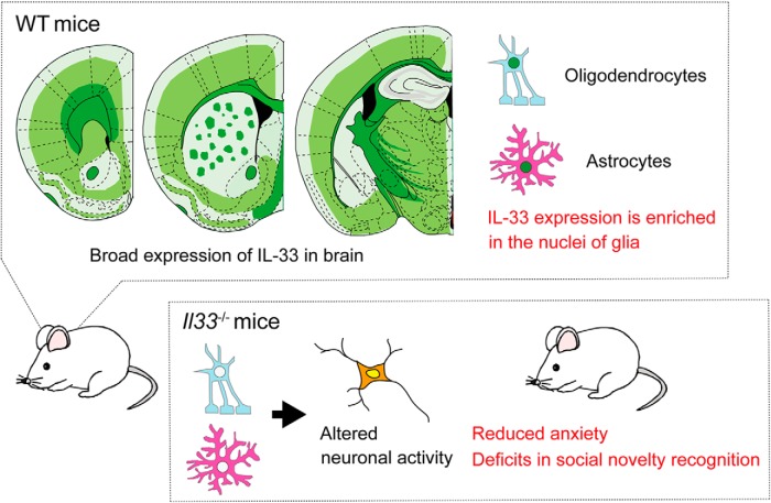
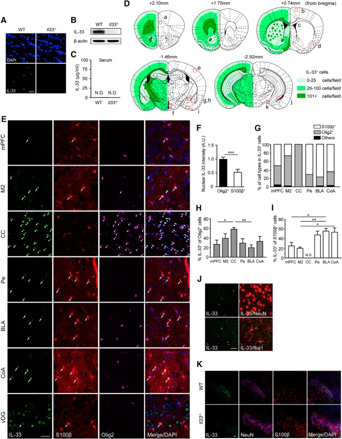
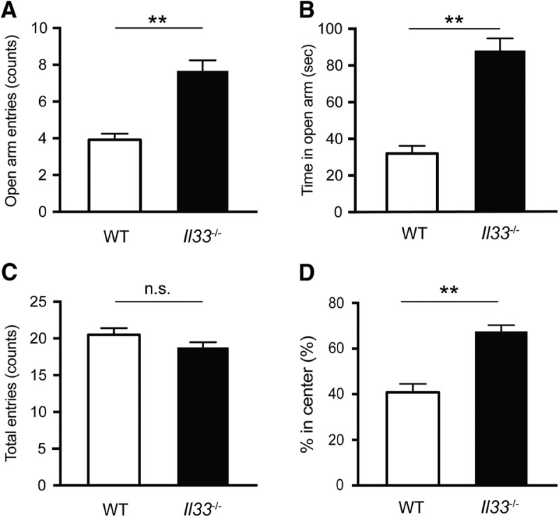
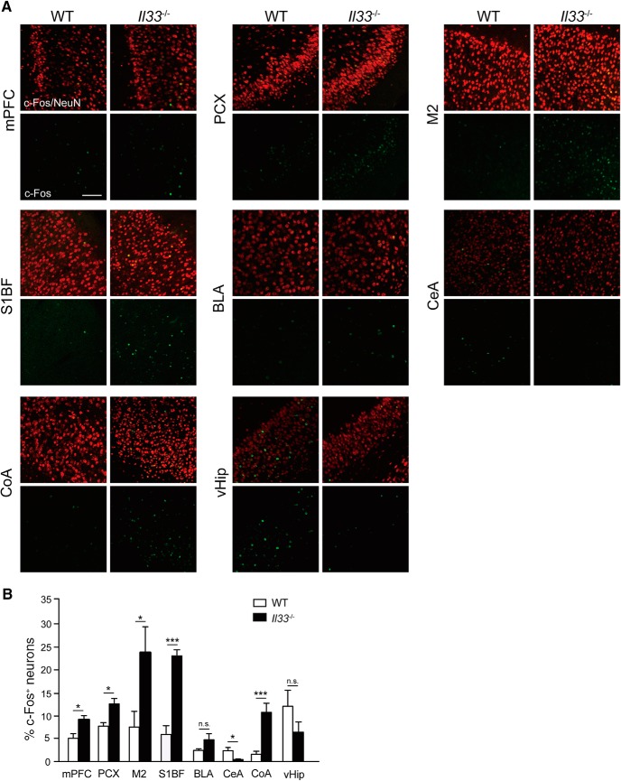
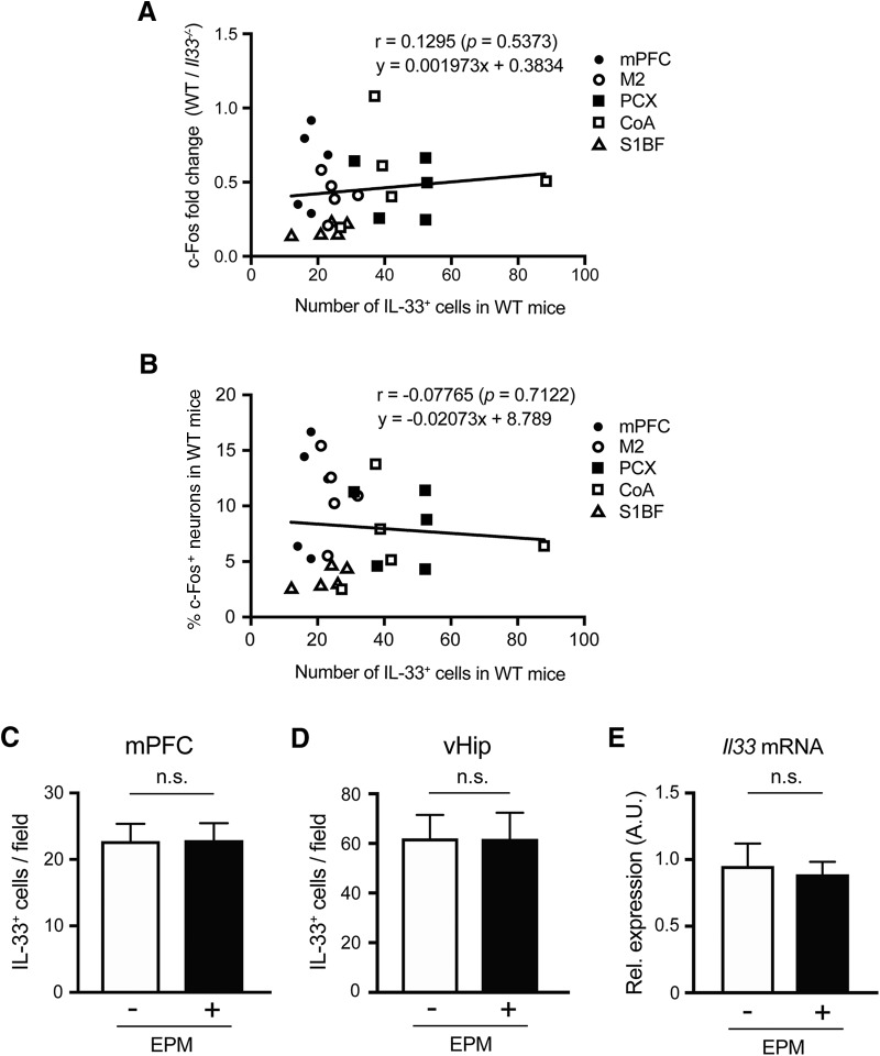
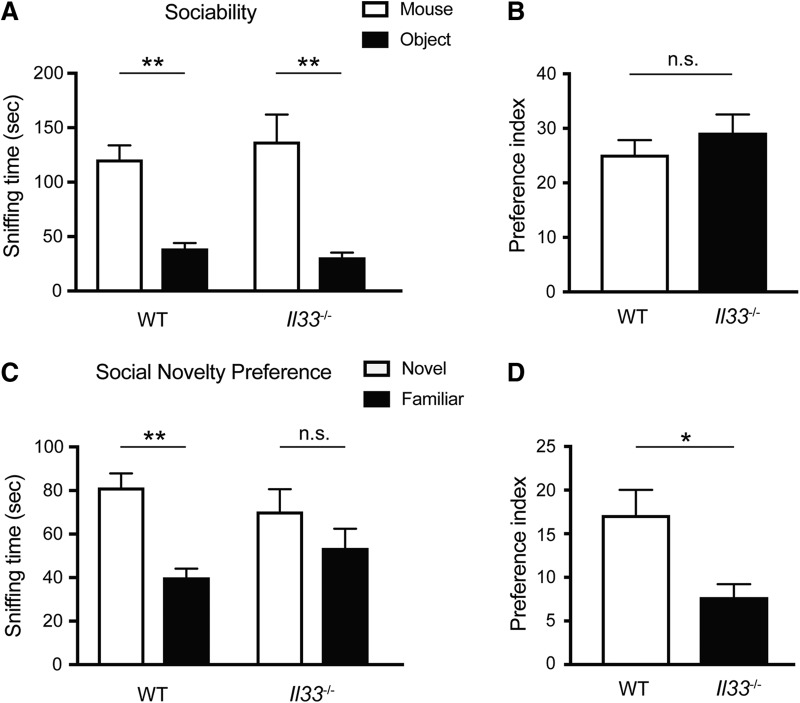
Interleukin (IL)-33 is a member of the IL-1 family of cytokines. IL-33 is expressed in nuclei and secreted as alarmin upon cellular damage to deliver a danger signal to the surrounding cells. Previous studies showed that IL-33 is expressed in the brain and that it is involved in neuroinflammatory and neurodegenerative processes in both humans and rodents. Nevertheless, the role of IL-33 in physiological brain function and behavior remains unclear. Here, we have investigated the behaviors of mice lacking IL-33 (Il33-/- mice). IL-33 is constitutively expressed throughout the adult mouse brain, mainly in oligodendrocyte-lineage cells and astrocytes. Notably, Il33-/- mice exhibited reduced anxiety-like behaviors in the elevated plus maze (EPM) and the open field test (OFT), as well as deficits in social novelty recognition, despite their intact sociability, in the three-chamber social interaction test. The immunoreactivity of c-Fos proteins, an indicator of neuronal activity, was altered in several brain regions implicated in anxiety-related behaviors, such as the medial prefrontal cortex (mPFC), amygdala, and piriform cortex (PCX), in Il33-/- mice after the EPM. Altered c-Fos immunoreactivity in Il33-/- mice was not correlated with IL-33 expression in wild-type (WT) mice nor was IL-33 expression affected by the EPM in WT mice. Thus, our study has revealed that Il33-/- mice exhibit multiple behavioral deficits, such as reduced anxiety and impaired social recognition. Our findings also indicate that IL-33 may regulate the development and/or maturation of neuronal circuits, rather than control neuronal activities in adult brains.

Keywords: IL-33; anxiety; astrocytes; cytokines; oligodendrocytes; social behavior.


Figures

None
Graphical abstract
Figure 1.
Figure 1.
IL-33 expression patterns in the adult mouse brain. A, Validation of specificity of IL-33 immunostaining using brain sections (CC) from WT and Il33 −/− mice. B, Western blot analysis of IL-33 expression in the cortex from WT and Il33 −/− mice. β-actin was used as an internal control for protein loading. C, Absence of IL-33 proteins in the peripheral blood of WT mice. The levels of serum IL-33 proteins were below the detection threshold of ELISA assay (15.6 pg/ml). N.D., not detected. D, Schematic illustration of the distribution of IL-33-expressing cells. The number of IL-33-expressing cells was quantified at 20× under a fluorescence microscope. Light, medium, and dark green correspond to 0–25, 26–100, and 101+ cells per field, respectively. Small red boxes indicate the area analyzed in E–K and Figs. 3, 4A–D : a, mPFC; b, secondary motor cortex (M2); c, CC; d, PCX; e, primary S1BF; f, Pe; g, CeA; h, BLA; i, CoA; j, ventral hippocampus (vHip); k, vDG. E, Representative pictures of IL-33 expression in several brain regions. Arrows, IL-33+ astrocytes (S100β+ cells); arrowheads, IL-33+ oligodendrocyte-lineage cells (Olig2+ cells). F, The average nuclear intensity of IL-33 between Olig2+ cells and S100β+ cells in the M2. A.U., arbitrary unit. G, Percentages of S100β+, Olig2+, and other cells among IL-33+ cells in each brain region. H, Comparison of IL-33 expression in Olig2+ cells across brain regions. I, Comparison of IL-33 expression in S100β+ cells across brain regions. We also observed Olig2+ and S100β+ cells, but these cells are not included in this graph. J, No colocalization of IL-33 signals to neurons (NeuN+ cells) or microglia (Iba1+ cells) in most brain regions. Representative pictures of the M2 are shown. K, IL-33 colocalization to neurons in the granular layer of vDG. Scale bar, 30 µm. Each bar represents mean ± SEM; *p < 0.05, **p < 0.01 (Student’s t test, one-way ANOVA with post hoc Tukey’s test, and Kruskal–Wallis test with post hoc Dunnett’s test; see Table 2 for the detail of statistical analysis).
Figure 2.
Figure 2.
Reduced anxiety-like behavior in Il33 −/− mice. A, Increased open arm entries into the EPM. B, Increased time spent in the open arms in the EPM. C, No difference in total entries between WT and Il33 −/− mice in the EPM. WT mice, n = 12; Il33 −/− mice, n = 9. D, Increased time spent in the center during the OFT. WT mice, n = 9; Il33 −/− mice, n = 7. Each bar represents mean ± SEM; n.s, not significant; **p < 0.01 (Student’s t test; see Table 2 for the detail of statistical analysis).
Figure 3.
Figure 3.
Altered c-Fos immunoreactivity in brain regions related to anxiety in Il33 −/− mice. A, Representative images of c-Fos expression in NeuN+ neurons. Location of each brain region is indicated in Fig. 1D [a, mPFC; b, M2; d, PCX; e, S1BF; g, CeA; h, BLA; i, CoA; k, ventral hippocampus (vHip)]. B, Quantification of c-Fos expression in various brain regions. Percentages of c-Fos-expressing neurons among all neurons (NeuN+ cells) were calculated and compared between WT and Il33 −/− mice (n = 3-4). Scale bar, 100 µm. Each bar represents mean ± SEM; n.s., not significant; *p < 0.05, **p < 0.01 (Student’s t test; see Table 2 for the detail of statistical analysis).
Figure 4.
Figure 4.
Relationship between IL-33 expression and c-Fos immunoreactivity. A, Correlation between the number of IL-33+ cells per field in WT mice and fold change of % c-Fos+ neurons (NeuN+ cells) per field in WT mice normalized with average % c-Fos+ neurons per field in Il33 −/− mice. Each dot refers to the brain region of an individual mouse (WT mice, n = 5; Il33−/− mice, n = 5). B, Correlation between the % c-Fos+ neurons per field in WT mice and the number of IL-33+ cells per field in WT mice based on c-Fos elevated brain regions in Il33−/− mice after EPM. Each dot refers to the brain region of an individual mouse. C, D, No difference in IL-33 expression in the mPFC and ventral hippocampus (vHip) between WT mice with and without the EPM (EPM+ and EPM). E, No difference in the Il33 mRNA expression in the fontal cortex between EPM+ and EPM-. EPM+ n = 4, EPM n = 4. A.U., arbitrary unit. Each bar represents mean ± SEM; n.s., not significant (Pearson correlation, Student’s t test; see Table 1 for the detail of statistical analysis).
Figure 5.
Figure 5.
Altered social behaviors in Il33 −/− mice. A, No significant difference in sociability between WT and Il33 −/− mice. (Fgenotype x chamber(1,22) = 0.9178, p = 0.3485; Fgenotype(1,22) = 0.09669, p = 0.7588; FChamber(1,22) = 54.44, p < 0.001). Both WT and Il33 −/− mice preferred mice to objects (**p < 0.01, post hoc Sidak test). B, Preference index data for sociability behaviors (p = 0.3587, Student’s t test). C, Reduced preference to novel mice in Il33 −/− mice. WT and Il33 −/− mice differed significantly in preference trial (Fgenotype x chamber(1,22) = 4.681, p < 0.05; Fgenotype(1,22) = 0.00224, p = 0.8824; FChamber(1,22) = 26.34; p < 0.0001). Only WT mice showed a significant preference to novel mice (**p < 0.01, post hoc Sidak test). D, Preference index data for social novelty preference. Il33 −/− group showed a significantly lower preference index than the WT group (*p < 0.05, Student’s t test). Each bar represents mean ± SEM; n.s., not significant. See Table 2 for the detail of statistical analysis.

References

    1. Ali S, Mohs A, Thomas M, Klare J, Ross R, Schmitz ML, Martin MU (2011) The dual function cytokine IL-33 interacts with the transcription factor NF-κB to dampen NF-κB-stimulated gene transcription. J Immunol 187:1609–1616. 10.4049/jimmunol.1003080 - DOI - PubMed
    1. Aston C, Jiang L, Sokolov BP (2005) Transcriptional profiling reveals evidence for signaling and oligodendroglial abnormalities in the temporal cortex from patients with major depressive disorder. Mol Psychiatry 10:309–322. 10.1038/sj.mp.4001565 - DOI - PubMed
    1. Banasr M, Duman RS (2008) Glial loss in the prefrontal cortex is sufficient to induce depressive-like behaviors. Biol Psychiatry 64:863–870. 10.1016/j.biopsych.2008.06.008 - DOI - PMC - PubMed
    1. Ben-Hamo M, Larson TA, Duge LS, Sikkema C, Wilkinson CW, de la Iglesia HO, González MM (2016) Circadian forced desynchrony of the master clock leads to phenotypic manifestation of depression in rats. eNeuro 3. - PMC - PubMed
    1. Calhoon GG, Tye KM (2015) Resolving the neural circuits of anxiety. Nat Neurosci 18:1394–1404. 10.1038/nn.4101 - DOI - PMC - PubMed

Publication types

LinkOut - more resources