. 2018 Jun;169(2):231-241.
doi: 10.1007/s10549-018-4664-7. Epub 2018 Jan 29.

The PDGF pathway in breast cancer is linked to tumour aggressiveness, triple-negative subtype and early recurrence

Affiliations

The PDGF pathway in breast cancer is linked to tumour aggressiveness, triple-negative subtype and early recurrence

Sara Jansson et al. Breast Cancer Res Treat. 2018 Jun.

Abstract

Purpose: The platelet-derived growth factor (PDGF) signalling pathway is often dysregulated in cancer and PDGF-receptor expression has been linked to unfavourable prognostic factors in breast cancer (e.g. ER negativity, high Ki67 and high grade). This study aimed to evaluate the expression of PDGFRα, PDGFRβ and ligand PDGF-CC in breast cancer in relation to molecular subtypes and prognosis.

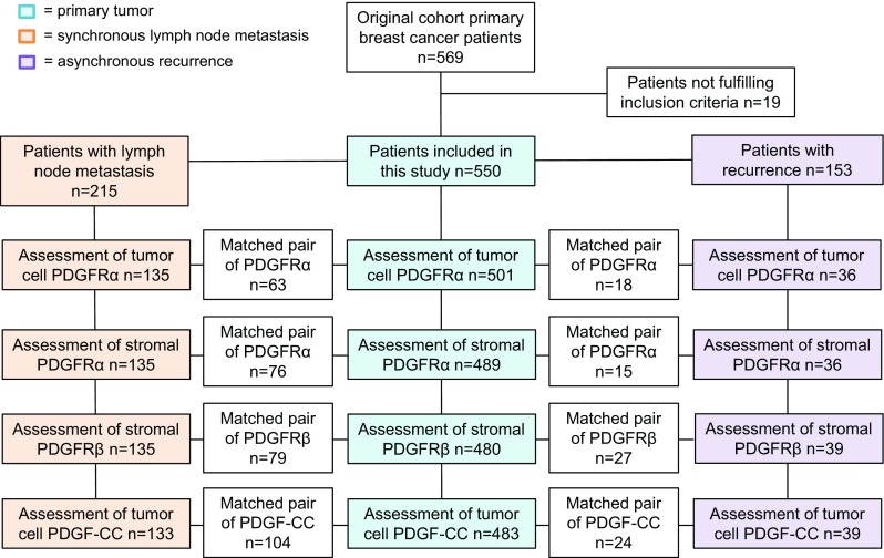
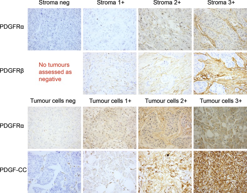
Methods: Protein expression of tumour and/or stromal cell PDGFRα, PDGFRβ and PDGF-CC was evaluated in primary tumours (N = 489), synchronous lymph node metastases (N = 135) and asynchronous recurrences (N = 39) using immunohistochemistry in a prospectively maintained cohort of primary breast cancer patients included during 1999-2003. Distant recurrence-free interval (DRFi) was the primary end-point.

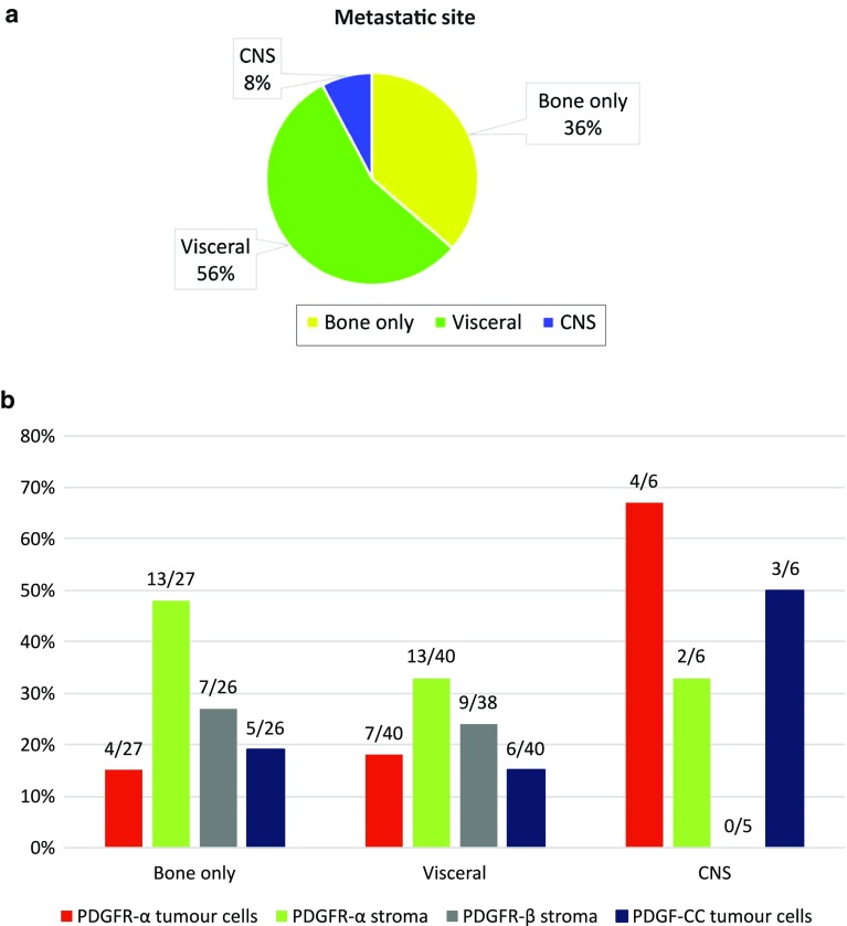
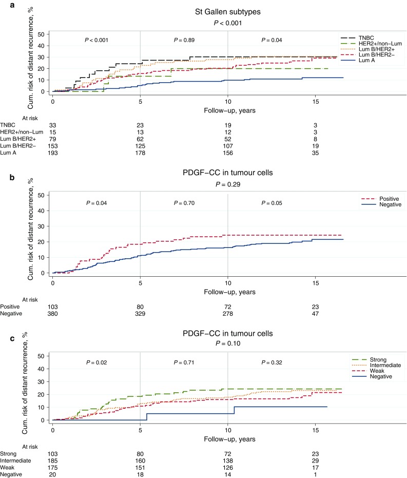
Results: High expression of all investigated PDGF family members correlated to increasing Nottingham histopathological grade and high Ki67. Tumour cells displayed high expression of PDGFRα in 20%, and PDGF-CC in 21% of primary tumours, which correlated with the triple-negative subtype (TNBC). Patients with high PDGF-CC had inferior prognosis (P = 0.04) in terms of 5-year DRFi, whereas PDGFRα was up-regulated in lymph node metastasis and recurrences compared to primary tumours. High primary tumour PDGFRα was associated with increased risk of central nervous system (CNS) recurrence.

Conclusions: High PDGFRα and PDGF-CC expression were linked to breast cancer with an aggressive biological phenotype, e.g. the TNBC subtype, and high PDGF-CC increased the risk of 5-year distant recurrence. Tumour cell PDGFRα was significantly up-regulated in lymph node metastases and asynchronous recurrences. Our findings support an active role of the PDGF signalling pathway in tumour progression.

Keywords: Breast cancer; Platelet-derived growth factor receptor; Platelet-derived growth factor-CC; Targeted therapy; Triple-negative breast cancer; Tyrosine kinase receptor.


Conflict of interest statement

The authors declare that they have no conflict of interest.

Figures

Fig. 1
Fig. 1
Flowchart of patient cohort and biomarker expression in primary tumour, synchronous lymph node metastases and asynchronous recurrences. Due to limited remaining tissue material, synchronous lymph node metastasis and asynchronous recurrences were only evaluable in 135 and 39 patients, respectively. Boxes are inserted into the flowchart displaying information on matched pairs, i.e. numbers of primary tumours and nodes, and primary tumours and recurrences displaying identical scoring of each marker, respectively (positive–positive or negative–negative)
Fig. 2
Fig. 2
Examples of IHC stainings for the members of the PDGF family. PDGFRα in stromal cells (1st row), PDGFRβ in stromal cells (2nd row), PDGFRα in tumour cells (3rd row) and PDGF-CC in tumour cells (4th row). Original magnification ×40
Fig. 3
Fig. 3
a Overview of primary metastatic site at time of recurrence. b Relation between primary tumour PDGF expression (receptors α, β or ligand –CC) and site of distant recurrence
Fig. 4
Fig. 4
ac Kaplan–Meier survival curves showing DRFi (years) in relation to St Gallen molecular subtypes (a), expression of PDGF-CC in tumour cells dichotomized into positive versus negative (b), and staining intensity of PDGF-CC ranging from 0 (negative) to 3 (strong) (c). P values from log rank test and log rank linear trends for factor levels

References

    1. Demoulin JB, Essaghir A. PDGF receptor signaling networks in normal and cancer cells. Cytokine Growth Factor Rev. 2014;25(3):273–283. doi: 10.1016/j.cytogfr.2014.03.003. - DOI - PubMed
    1. Nazarenko I, Hede SM, He X, Hedren A, Thompson J, Lindstrom MS, Nister M. PDGF and PDGF receptors in glioma. Upsala J Med Sci. 2012;117(2):99–112. doi: 10.3109/03009734.2012.665097. - DOI - PMC - PubMed
    1. Heinrich MC, Corless CL, Duensing A, McGreevey L, Chen CJ, Joseph N, Singer S, Griffith DJ, Haley A, Town A, Demetri GD, Fletcher CD, Fletcher JA. PDGFRA activating mutations in gastrointestinal stromal tumors. Science. 2003;299(5607):708–710. doi: 10.1126/science.1079666. - DOI - PubMed
    1. Golub TR, Barker GF, Lovett M, Gilliland DG. Fusion of PDGF receptor beta to a novel ets-like gene, tel, in chronic myelomonocytic leukemia with t(5;12) chromosomal translocation. Cell. 1994;77(2):307–316. doi: 10.1016/0092-8674(94)90322-0. - DOI - PubMed
    1. Ehnman M, Ostman A. Therapeutic targeting of platelet-derived growth factor receptors in solid tumors. Expert Opin Investig Drugs. 2014;23(2):211–226. doi: 10.1517/13543784.2014.847086. - DOI - PubMed

MeSH terms

Substances

LinkOut - more resources