

Cornea and anterior eye assessment with slit lamp biomicroscopy, specular microscopy, confocal microscopy, and ultrasound biomicroscopy
- PMID: 29380757
- PMCID: PMC5819094
- DOI: 10.4103/ijo.IJO_649_17
Cornea and anterior eye assessment with slit lamp biomicroscopy, specular microscopy, confocal microscopy, and ultrasound biomicroscopy
Abstract
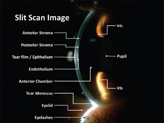
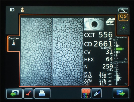
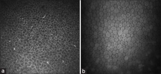
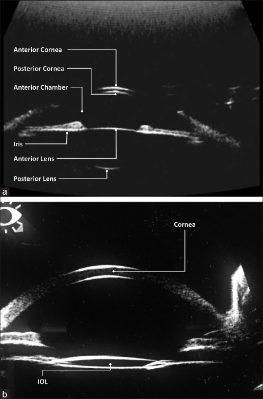
Current corneal assessment technologies make the process of corneal evaluation extremely fast and simple, and several devices and technologies show signs that help in identification of different diseases thereby, helping in diagnosis, management, and follow-up of patients. The purpose of this review is to present and update readers on the evaluation of cornea and ocular surface. This first part reviews a description of slit lamp biomicroscopy (SLB), endothelial specular microscopy, confocal microscopy, and ultrasound biomicroscopy examination techniques and the second part describes the corneal topography and tomography, providing up-to-date information on the clinical recommendations of these techniques in eye care practice. Although the SLB is a traditional technique, it is of paramount importance in clinical diagnosis and compulsory when an eye test is conducted in primary or specialist eye care practice. Different techniques allow the early diagnosis of many diseases, especially when clinical signs have not yet become apparent and visible with SLB. These techniques also allow for patient follow-up in several clinical conditions or diseases, facilitating clinical decisions and improving knowledge regarding the corneal anatomy.
Conflict of interest statement
There are no conflicts of interest.
Figures
References
-
- Rio-Cristobal A, Martin R. Corneal assessment technologies: Current status. Surv Ophthalmol. 2014;59:599–614. - PubMed
-
- Brody J, Waller S, Wagoner M. Corneal topography: History, technique, and clinical uses. Int Ophthalmol Clin. 1994;34:197–207. - PubMed
-
- Fleming JB, Semes LP. Benjamin WJ. Borish's Clinical Refraction. 2nd ed. St Louis, Missouri (USA): Butterworth-Heinemann-Elsevier; 2006. Anterior segment evaluation.
-
- Efron N. Contact Lens Complications. 3rd ed. New York (USA): Elsevier Saunders; 2012.
-
- Efron N. Contact lens-induced changes in the anterior eye as observed in vivo with the confocal microscope. Prog Retin Eye Res. 2007;26:398–436. - PubMed
Publication types
MeSH terms
LinkOut - more resources
Full Text Sources
Other Literature Sources
Medical
