

High-resolution optical coherence tomography in a case of descemetocele managed with amniotic membrane transplantation
- PMID: 29380791
- PMCID: PMC5819128
- DOI: 10.4103/ijo.IJO_697_17
High-resolution optical coherence tomography in a case of descemetocele managed with amniotic membrane transplantation
Abstract
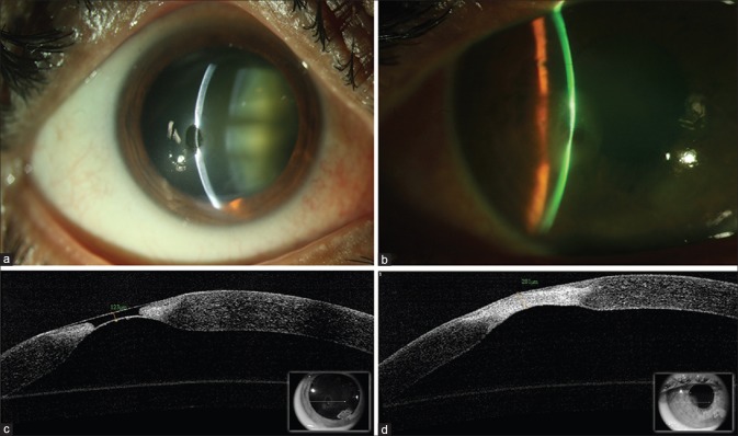
Amniotic membrane transplantation is a useful in the management of corneal melts and descemetocele. We describe high-resolution anterior segment optical coherence tomography (OCT) (Optovue) in a patient with descemetocele who was managed surgically with amniotic membrane transplantation. A 60-year-old female presented with a corneal melt in the right eye. She was a case of rheumatoid arthritis and was on systemic treatment with immunomodulators. Slit lamp examination revealed a severe thinning paracentrally. High-resolution OCT was performed at the site of descemetocele. She underwent amniotic membrane transplantation using fibrin glue and bandage contact lens application. At 6 weeks postoperative, the bandage contact lens was removed. The area of thinning healed with scarring. OCT at the healed site revealed stable surface and an increase in stromal thickness to 281 μ this case describes the utility of amniotic membrane in the healing of sterile corneal melts by providing tectonic support and its integration in the stroma. The stromal healing and increased thickness at the site of descemetocele could be delineated on high-resolution OCT imaging.
Conflict of interest statement
There are no conflicts of interest.
Figures
References
-
- Tong L, Thumboo J, Tan YK, Wong TY, Albani S. The eye: A window of opportunity in rheumatoid arthritis? Nat Rev Rheumatol. 2014;10:552–60. - PubMed
-
- Bernauer W, Ficker LA, Watson PG, Dart JK. The management of corneal perforations associated with rheumatoid arthritis. An analysis of 32 eyes. Ophthalmology. 1995;102:1325–37. - PubMed
-
- Solomon A, Meller D, Prabhasawat P, John T, Espana EM, Steuhl KP, et al. Amniotic membrane grafts for nontraumatic corneal perforations, descemetoceles, and deep ulcers. Ophthalmology. 2002;109:694–703. - PubMed
-
- Kim HK, Park HS. Fibrin glue-assisted augmented amniotic membrane transplantation for the treatment of large noninfectious corneal perforations. Cornea. 2009;28:170–6. - PubMed
-
- Lee SH, Tseng SC. Amniotic membrane transplantation for persistent epithelial defects with ulceration. Am J Ophthalmol. 1997;123:303–12. - PubMed
Publication types
MeSH terms
LinkOut - more resources
Full Text Sources
Medical
Research Materials
