. 2018 Apr;108(1):90-100.
doi: 10.1111/mmi.13918. Epub 2018 Feb 19.

Rapid evolution of a bacterial iron acquisition system

Affiliations

Rapid evolution of a bacterial iron acquisition system

Anushila Chatterjee et al. Mol Microbiol. 2018 Apr.

Abstract

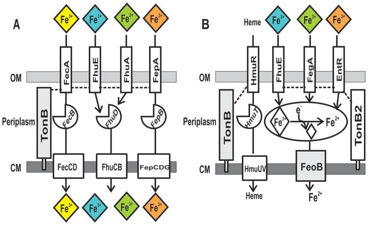
Under iron limitation, bacteria scavenge ferric (Fe3+ ) iron bound to siderophores or other chelates from the environment to fulfill their nutritional requirement. In gram-negative bacteria, the siderophore uptake system prototype consists of an outer membrane transporter, a periplasmic binding protein and a cytoplasmic membrane transporter, each specific for a single ferric siderophore or siderophore family. Here, we show that spontaneous single gain-of-function missense mutations in outer membrane transporter genes of Bradyrhizobium japonicum were sufficient to confer on cells the ability to use synthetic or natural iron siderophores, suggesting that selectivity is limited primarily to the outer membrane and can be readily modified. Moreover, growth on natural or synthetic chelators required the cytoplasmic membrane ferrous (Fe2+ ) iron transporter FeoB, suggesting that iron is both dissociated from the chelate and reduced to the ferrous form within the periplasm prior to cytoplasmic entry. The data suggest rapid adaptation to environmental iron by facile mutation of selective outer membrane transporter genes and by non-selective uptake components that do not require mutation to accommodate new iron sources.


Figures

FIGURE 1
FIGURE 1. Dominant gain-of-function mutations in siderophore outer membrane transporter genes rescue growth on the synthetic iron chelator EDDHA
Cells were serial diluted, and 5 μl aliquots were spotted onto plates 20 μM FeCl3 (A), 100 μM EDDHA (B), 100 μM EDDHA plus 20 μM FeCl3 (C), 20 μM DTPA (D), 20 μM DTPA and 100 μM EDDHA (E) or 20 μM DTPA with 20 μM FeCl3 (F). The strains entR(L272P), entR(T437P) and fegA(L497Q) represent the isogenic siderophore transporter mutants reconstructed in the wild type background. Strains entR(L272P)+, entR(T437P)+ and fegA(L497Q)+ denote the partial diploids harboring the wild type copy and the designated mutated copy of the transporter, integrated in the wild type genome.
FIGURE 2
FIGURE 2. Requirement of tonB2 for growth on natural siderophores or EDDHA
Serial dilution and spotting on plates of modified GSY media containing 10 μM heme (A), 100 μM EDDHA (B), 4 μM ferrichrome (C), 4 μM enterobactin (D) or 25 μM desferrioxamine (E). The tonB2 and entR(L272P) tonB2 strains have an in-frame deletion in the tonB2 (blr3908) gene in the wild type and entR(L272P) background respectively. The fegA, entR and fhuE strains are defective in the respective ferric-siderophore transporters.
FIGURE 3
FIGURE 3. Uptake of siderophore-mediated 59Fe3+
Uptake of 59Fe3+- siderophores was measured in the cells of the wild type, tonB2 and feoB strains along with the respective siderophore transporter mutant entR, fegA or fhuE. Cells were suspended in uptake buffer containing 4 μM enterobactin (A), 4 μM ferrichrome (B) or 25 μM desferrioxamine (C). At time 0, 25 nM 59Fe3+ was added to the cell suspension, and aliquots were subsequently collected at 0, 10, 50 minutes to measure counts. Each time point is the average of three biological replicates ± S.D. (error bars) after subtracting the 0 minute reading.
FIGURE 4
FIGURE 4. Gain-of-function mutations in the heme transporter gene hmuR suppress the growth and Fe3+ uptake phenotypes of an fhuE mutant on desferrioxamine
(A) Growth of mutants on desferrioxamine. Cells were serially diluted, and 5 μl aliquots were spotted onto plates containing 20 μM FeCl3 (a), 25 μM desferrioxamine (b) or 25 μM desferrioxamine plus 20 μM FeCl3 (c). Strains with a + sign denote partial diploids with both the wild type and the mutated hmuR allele integrated into the genome. (B) 59Fe3+- desferrioxamine uptake. Cells of the wild type (open square), fhuE (closed square), fhuE hmuR(W351R) (open triangle), fhuE hmuR(Δ182-193) (open circle) and fhuE hmuR (Δ403-425) (closed circle) strains were suspended in uptake buffer containing 25 μM desferrioxamine (a) or 20 mM sodium-citrate (b). At time 0, 50 nM 59Fe3+ was added to the cell suspension, and aliquots were subsequently taken at various time points and counted. Each time point is the average of three biological replicate samples ± S.D. (error bars).
FIGURE 5
FIGURE 5. FeoB is essential for the utilization of natural and synthetic iron chelates
(A) Growth on natural xenosiderophores. Serial dilution and spotting of cells grown in liquid GSY media with 100nM heme was performed on modified GSY media plates containing 10 μM heme (a), no added iron (b), 20 μM FeCl3 (c), 4 μM ferrichrome (d), 4 μM enterobactin (e) or 25 μM desferrioxamine (f). The fegA, entR and fhuE strains have defects in the ferricrhome, enterobactin and desferrioxamine transporter respectively. (B) Growth on EDDHA. Cells were grown in liquid GSY media with 100 nM heme, serially diluted and spotted on modified GSY media containing 10 μM heme (a), 100 μM EDDHA (b) or 100 μM EDDHA plus 10 μM heme (c). The entR(L272P) feoB strain has in-frame deletion in the feoB gene in the entR(L272P) background.
FIGURE 6
FIGURE 6. B. japonicum is resistant to albomycin compared with E. coli cells
Cells were serially diluted, and 5 μl aliquots were spotted onto plates in the presence or absence of 2 μM albomycin. (A) E. coli cells. (B) B. japonicum strain 110 (Bj) or B. japonicum cells harboring the E. coli ferrichrome transport genes fhuCDB
FIGURE 7
FIGURE 7. Comparison of ferric-chelate acquisition systems between E. coli and B. japonicum
(A) In E.coli, outer membrane transporters FecA, FepA, FhuE, FhuA import Fe3+ chelates of citrate, enterobactin, desferrioxamine and ferrichrome, respectively. Translocation of ferric-chelates across the outer membrane is dependent on TonB (auxiliary proteins ExbB and ExbD are not shown). (B) In B. japonicum, intact heme is transported into the cytoplasm through the TonB-dependent outer membrane heme transporter, HmuR. The outer membrane xenosiderophore transporters FhuE, FegA, and EntR import Fe3+ chelates of desferrioxamine, ferrichrome and enterobactin, respectively. These transporters depend on TonB2. In the periplasm, Fe3+-siderophore is likely reduced to Fe2+ iron, potentially causing its release from the chelator. A single inner membrane transporter, FeoB, is responsible for importing Fe2+ iron into the cytoplasm.

Similar articles

Cited by

References

    1. Alteri CJ, Hagan EC, Sivick KE, Smith SN, Mobley HLT. Mucosal immunization with iron receptor antigens protects against urinary tract infection. PLoS pathogens. 2009;5:e1000586. - PMC - PubMed
    1. Amarelle V, Koziol U, Rosconi F, Noya F, O’Brian MR, Fabiano E. A new small regulatory protein, HmuP, modulates haemin acquisition in Sinorhizobium meliloti. Microbiology. 2010;156:1873–1882. - PMC - PubMed
    1. Andrews SC, Robinson AK, Rodriguez-Quinones F. Bacterial iron homeostasis. FEMS Microbiol Rev. 2003;27:215–237. - PubMed
    1. Braun V, Günthner K, Hantke K, Zimmermann L. Intracellular activation of albomycin in Escherichia coli and Salmonella typhimurium. Journal of Bacteriology. 1983;156:308–315. - PMC - PubMed
    1. Braun V, Pramanik A, Gwinner T, Köberle M, Bohn E. Sideromycins: tools and antibiotics. BioMetals. 2009;22:3. - PMC - PubMed

Publication types

MeSH terms