

Smart Cell Culture Systems: Integration of Sensors and Actuators into Microphysiological Systems
- PMID: 29381325
- PMCID: PMC5959007
- DOI: 10.1021/acschembio.7b01029
Smart Cell Culture Systems: Integration of Sensors and Actuators into Microphysiological Systems
Abstract
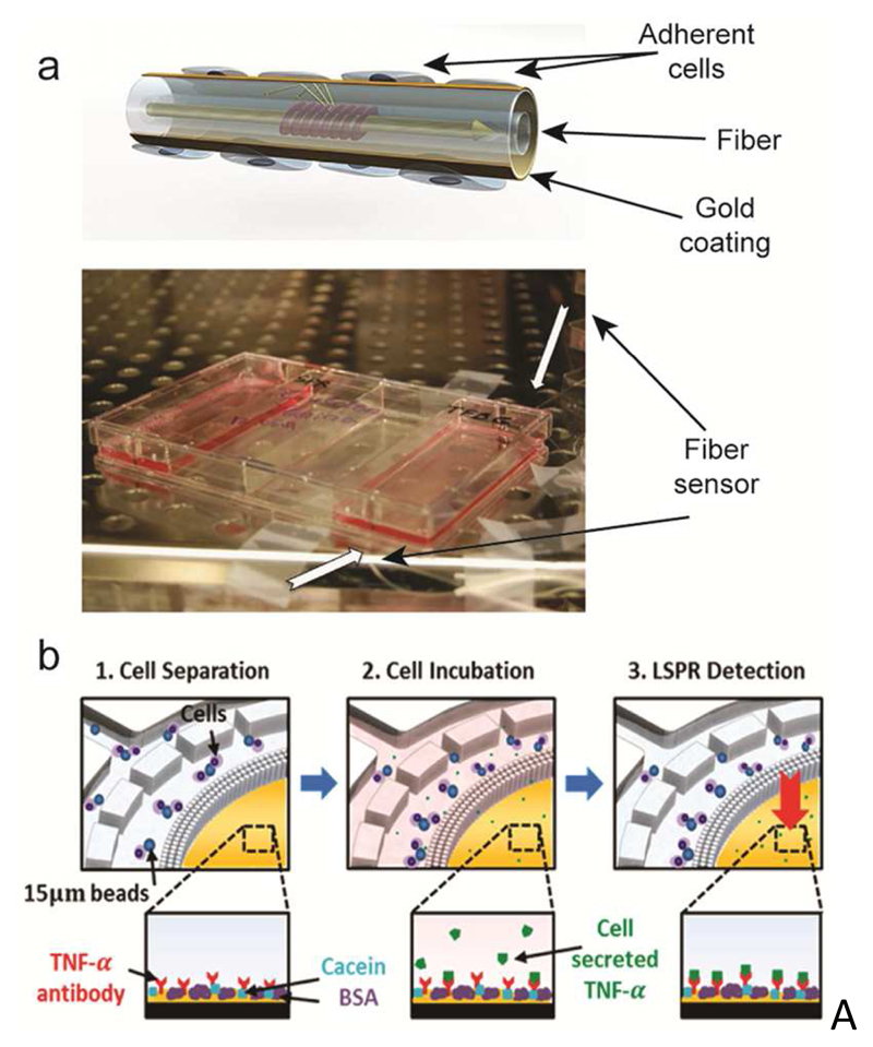
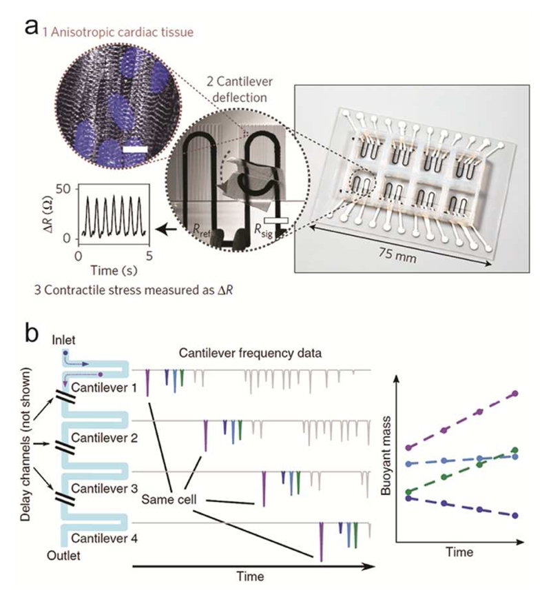
Technological advances in microfabrication techniques in combination with organotypic cell and tissue models have enabled the realization of microphysiological systems capable of recapitulating aspects of human physiology in vitro with great fidelity. Concurrently, a number of analysis techniques has been developed to probe and characterize these model systems. However, many assays are still performed off-line, which severely compromises the possibility of obtaining real-time information from the samples under examination, and which also limits the use of these platforms in high-throughput analysis. In this review, we focus on sensing and actuation schemes that have already been established or offer great potential to provide in situ detection or manipulation of relevant cell or tissue samples in microphysiological platforms. We will first describe methods that can be integrated in a straightforward way and that offer potential multiplexing and/or parallelization of sensing and actuation functions. These methods include electrical impedance spectroscopy, electrochemical biosensors, and the use of surface acoustic waves for manipulation and analysis of cells, tissue, and multicellular organisms. In the second part, we will describe two sensor approaches based on surface-plasmon resonance and mechanical resonators that have recently provided new characterization features for biological samples, although technological limitations for use in high-throughput applications still exist.
Figures
Similar articles
-
Advances in electrochemical-optical dual-mode biosensors for detection of environmental pathogens.Anal Methods. 2024 Feb 29;16(9):1306-1322. doi: 10.1039/d3ay02217j. Anal Methods. 2024. PMID: 38344759 Review.
-
Charge-based detection of small molecules by plasmonic-based electrochemical impedance microscopy.Anal Chem. 2013 Jul 16;85(14):6682-7. doi: 10.1021/ac400475z. Epub 2013 Jul 3. Anal Chem. 2013. PMID: 23815069
-
Electrochemically modulated surface plasmon waves for characterization and interrogation of DNA-based sensors.Analyst. 2024 Dec 2;149(24):5821-5831. doi: 10.1039/d4an01164c. Analyst. 2024. PMID: 39529568
-
In-Line Analysis of Organ-on-Chip Systems with Sensors: Integration, Fabrication, Challenges, and Potential.ACS Biomater Sci Eng. 2021 Jul 12;7(7):2926-2948. doi: 10.1021/acsbiomaterials.0c01110. Epub 2021 Jun 16. ACS Biomater Sci Eng. 2021. PMID: 34133114 Free PMC article.
-
Advancements in SPR biosensing technology: An overview of recent trends in smart layers design, multiplexing concepts, continuous monitoring and in vivo sensing.Anal Chim Acta. 2020 Apr 1;1104:10-27. doi: 10.1016/j.aca.2019.12.067. Epub 2019 Dec 27. Anal Chim Acta. 2020. PMID: 32106939 Review.
Cited by
-
Electrochemical Sensing in 3D Cell Culture Models: New Tools for Developing Better Cancer Diagnostics and Treatments.Cancers (Basel). 2021 Mar 18;13(6):1381. doi: 10.3390/cancers13061381. Cancers (Basel). 2021. PMID: 33803738 Free PMC article. Review.
-
dCas9-VPR-mediated transcriptional activation of functionally equivalent genes for gene therapy.Nat Protoc. 2022 Mar;17(3):781-818. doi: 10.1038/s41596-021-00666-3. Epub 2022 Feb 7. Nat Protoc. 2022. PMID: 35132255 Review.
-
Schistosomiasis Drug Discovery in the Era of Automation and Artificial Intelligence.Front Immunol. 2021 May 31;12:642383. doi: 10.3389/fimmu.2021.642383. eCollection 2021. Front Immunol. 2021. PMID: 34135888 Free PMC article. Review.
-
Humanized brain organoids-on-chip integrated with sensors for screening neuronal activity and neurotoxicity.Mikrochim Acta. 2024 Jan 3;191(1):71. doi: 10.1007/s00604-023-06165-4. Mikrochim Acta. 2024. PMID: 38168828 Review.
-
Gas Partial Pressure in Cultured Cells: Patho-Physiological Importance and Methodological Approaches.Front Physiol. 2018 Dec 13;9:1803. doi: 10.3389/fphys.2018.01803. eCollection 2018. Front Physiol. 2018. PMID: 30618815 Free PMC article. Review.
References
-
- Fatehullah A, Tan SH, Barker N. Organoids as an in vitro model of human development and disease. Nat Cell Biol. 2016;18:246–254. - PubMed
-
- van Duinen V, Trietsch SJ, Joore J, Vulto P, Hankemeier T. Microfluidic 3D cell culture: From tools to tissue models. Curr Opin Biotechnol. 2015;35:118–126. - PubMed
-
- Giridharan GA, Nguyen M-D, Estrada R, Parichehreh V, Hamid T, Ismahil MA, Prabhu SD, Sethu P. Microfluidic Cardiac Cell Culture Model (μCCCM) Anal Chem. 2010;82:7581–7587. - PubMed
-
- Korin N, Kanapathipillai M, Matthews BD, Crescente M, Brill A, Mammoto T, Ghosh K, Jurek S, Bencherif SA, Bhatta D, Coskun AU, et al. Shear-activated nanotherapeutics for drug targeting to obstructed blood vessels. Science. 2012;337:738–742. - PubMed
Publication types
MeSH terms
Grants and funding
LinkOut - more resources
Full Text Sources
Other Literature Sources
