

Rhinoscleroma pathogenesis: The type K3 capsule of Klebsiella rhinoscleromatis is a virulence factor not involved in Mikulicz cells formation
- PMID: 29381692
- PMCID: PMC5806929
- DOI: 10.1371/journal.pntd.0006201
Rhinoscleroma pathogenesis: The type K3 capsule of Klebsiella rhinoscleromatis is a virulence factor not involved in Mikulicz cells formation
Abstract
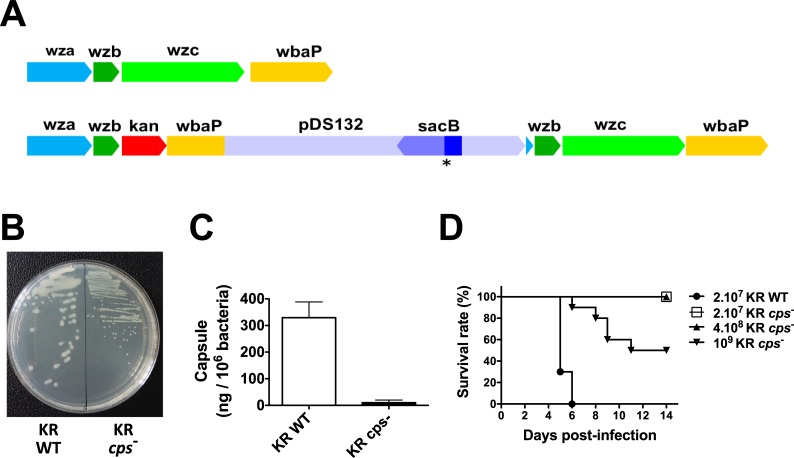
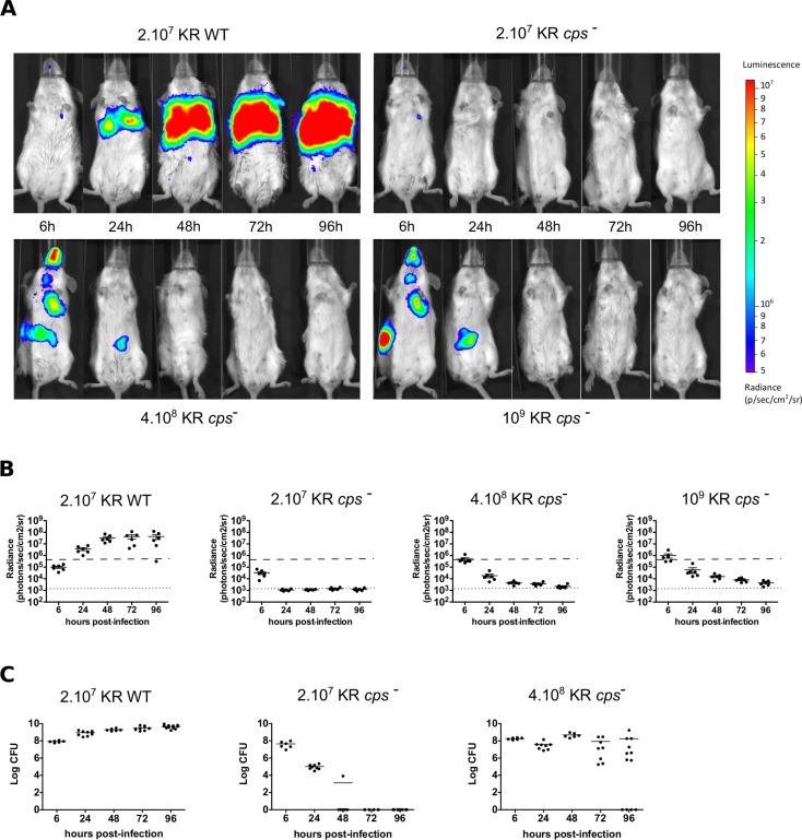
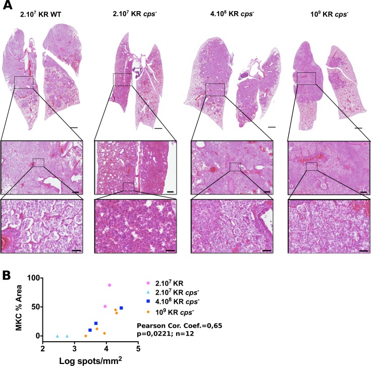
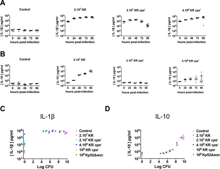
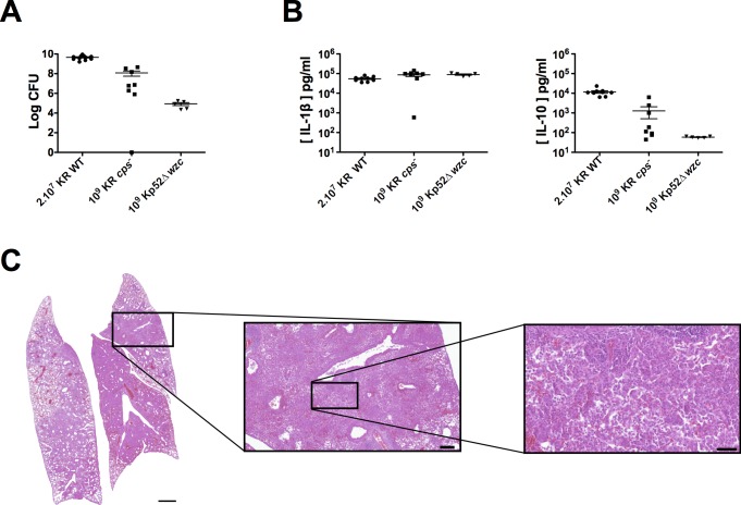
Rhinoscleroma is a human specific chronic granulomatous infection of the nose and upper airways caused by the Gram-negative bacterium Klebsiella pneumoniae subsp. rhinoscleromatis. Although considered a rare disease, it is endemic in low-income countries where hygienic conditions are poor. A hallmark of this pathology is the appearance of atypical foamy monocytes called Mikulicz cells. However, the pathogenesis of rhinoscleroma remains poorly investigated. Capsule polysaccharide (CPS) is a prominent virulence factor in bacteria. All K. rhinoscleromatis strains are of K3 serotype, suggesting that CPS can be an important driver of rhinoscleroma disease. In this study, we describe the creation of the first mutant of K. rhinoscleromatis, inactivated in its capsule export machinery. Using a murine model recapitulating the formation of Mikulicz cells in lungs, we observed that a K. rhinoscleromatis CPS mutant (KR cps-) is strongly attenuated and that mice infected with a high dose of KR cps- are still able to induce Mikulicz cells formation, unlike a K. pneumoniae capsule mutant, and to partially recapitulate the characteristic strong production of IL-10. Altogether, the results of this study show that CPS is a virulence factor of K. rhinoscleromatis not involved in the specific appearance of Mikulicz cells.
Conflict of interest statement
The authors have declared that no competing interests exist.
Figures
Similar articles
-
A novel murine model of rhinoscleroma identifies Mikulicz cells, the disease signature, as IL-10 dependent derivatives of inflammatory monocytes.EMBO Mol Med. 2013 Apr;5(4):516-30. doi: 10.1002/emmm.201202023. EMBO Mol Med. 2013. PMID: 23554169 Free PMC article.
-
PCR-based identification of Klebsiella pneumoniae subsp. rhinoscleromatis, the agent of rhinoscleroma.PLoS Negl Trop Dis. 2011 May;5(5):e1052. doi: 10.1371/journal.pntd.0001052. Epub 2011 May 24. PLoS Negl Trop Dis. 2011. PMID: 21629720 Free PMC article.
-
Experimental intravenous inoculation of Klebsiella rhinoscleromatis bacilli in albino rats: a histopathological and bacteriological study.Acta Otolaryngol. 2000 Mar;120(2):279-85. doi: 10.1080/000164800750001099. Acta Otolaryngol. 2000. PMID: 11603790
-
Klebsiella pneumoniae capsular polysaccharide: Mechanism in regulation of synthesis, virulence, and pathogenicity.Virulence. 2024 Dec;15(1):2439509. doi: 10.1080/21505594.2024.2439509. Epub 2024 Dec 13. Virulence. 2024. PMID: 39668724 Free PMC article. Review.
-
Social geography of Rhinoscleroma and qualitatively and quantitatively abnormal cell-mediated immunity.Infect Genet Evol. 2018 Aug;62:17-19. doi: 10.1016/j.meegid.2018.03.018. Epub 2018 Mar 22. Infect Genet Evol. 2018. PMID: 29578083 Review.
Cited by
-
Rhinoscleroma: Report of an Erratic Palatal Swelling.Contemp Clin Dent. 2018 Sep;9(Suppl 2):S365-S368. doi: 10.4103/ccd.ccd_282_18. Contemp Clin Dent. 2018. PMID: 30294174 Free PMC article.
-
Capsules and their traits shape phage susceptibility and plasmid conjugation efficiency.Nat Commun. 2024 Mar 6;15(1):2032. doi: 10.1038/s41467-024-46147-5. Nat Commun. 2024. PMID: 38448399 Free PMC article.
-
Resistance and virulence features of hypermucoviscous Klebsiella pneumoniae from bloodstream infections: Results of a nationwide Italian surveillance study.Front Microbiol. 2022 Aug 15;13:983294. doi: 10.3389/fmicb.2022.983294. eCollection 2022. Front Microbiol. 2022. PMID: 36204614 Free PMC article.
References
-
- Gaafar HA, Gaafar AH, Nour YA. Rhinoscleroma: an updated experience through the last 10 years. Acta Otolaryngol. 2011;131: 440–446. doi: 10.3109/00016489.2010.539264 - DOI - PubMed
-
- De Pontual L, Ovetchkine P, Rodriguez D, Grant A, Puel A, Bustamante J, et al. Rhinoscleroma: a French national retrospective study of epidemiological and clinical features. Clin Infect Dis. 2008;47: 1396–1402. doi: 10.1086/592966 - DOI - PubMed
-
- Jeffery J, Sulaiman S, Oothuman P, Vellayan S, Zainol-Ariffin P, Paramaswaran S, et al. Domiciliary cockroaches found in restaurants in five zones of Kuala Lumpur Federal Territory, peninsular Malaysia. Trop Biomed. 2011;29: 3309–3316. - PubMed
-
- Moges F, Eshetie S, Endris M, Huruy K, Muluye D, Feleke T, et al. Cockroaches as a Source of High Bacterial Pathogens with Multidrug Resistant Strains in Gondar Town, Ethiopia. Biomed Res Int. 2016;2016: 2825056 doi: 10.1155/2016/2825056 - DOI - PMC - PubMed
-
- Fielding BC, Mnabisa A, Gouws PA, Morris T. Antimicrobial-resistant Klebsiella species isolated from free-range chicken samples in an informal settlement. Arch Med Sci. 2012;8: 39–42. doi: 10.5114/aoms.2012.27278 - DOI - PMC - PubMed
Publication types
MeSH terms
Substances
LinkOut - more resources
Full Text Sources
Other Literature Sources
