Alteration in the Local and Global Functional Connectivity of Resting State Networks in Parkinson's Disease
- PMID: 29381889
- PMCID: PMC5790628
- DOI: 10.14802/jmd.17061
Alteration in the Local and Global Functional Connectivity of Resting State Networks in Parkinson's Disease
Abstract
Objective: Parkinson's disease (PD) is a neurodegenerative disorder that mainly leads to the impairment of patients' motor function, as well as of cognition, as it progresses. This study tried to investigate the impact of PD on the resting state functional connectivity of the default mode network (DMN), as well as of the entire brain.
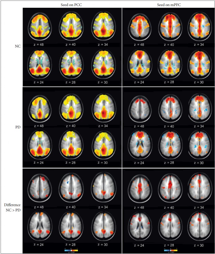
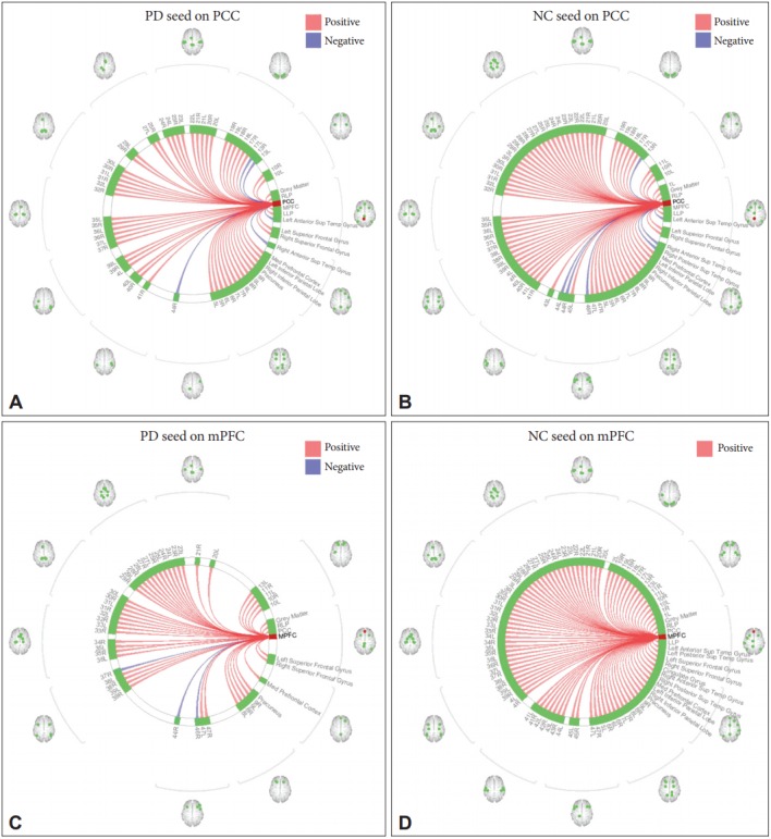
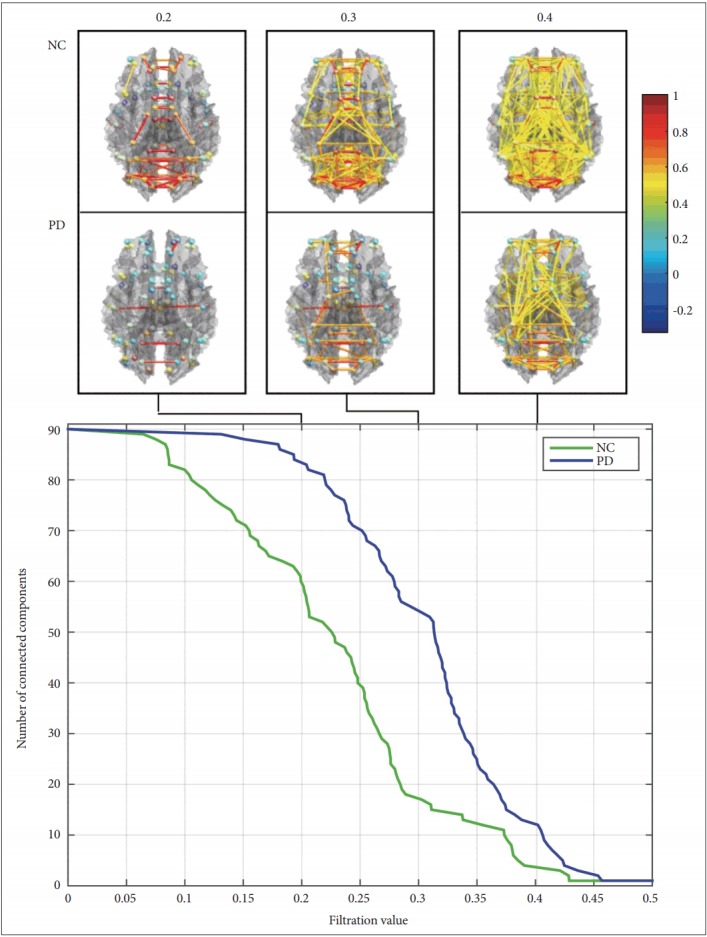
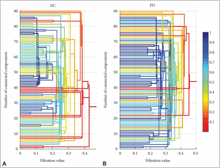
Methods: Sixty patients with PD were included and compared to 60 matched normal control (NC) subjects. For the local connectivity analysis, the resting state fMRI data were analyzed by seed-based correlation analyses, and then a novel persistent homology analysis was implemented to examine the connectivity from a global perspective.
Results: The functional connectivity of the DMN was decreased in the PD group compared to the NC, with a stronger difference in the medial prefrontal cortex. Moreover, the results of the persistent homology analysis indicated that the PD group had a more locally connected and less globally connected network compared to the NC.
Conclusion: Our findings suggest that the DMN is altered in PD, and persistent homology analysis, as a useful measure of the topological characteristics of the networks from a broader perspective, was able to identify changes in the large-scale functional organization of the patients' brain.
Keywords: Parkinson’s disease; default mode network; functional connectivity; persistent homology; resting state fMRI.
Conflict of interest statement
The authors have no financial conflicts of interest.
Figures
References
-
- Kalia LV, Lang AE. Parkinson’s disease. Lancet. 2015;386:896–912. - PubMed
-
- Rosazza C, Minati L. Resting-state brain networks: literature review and clinical applications. Neurol Sci. 2011;32:773–785. - PubMed
-
- Biswal BB, Van Kylen J, Hyde JS. Simultaneous assessment of flow and BOLD signals in resting-state functional connectivity maps. NMR Biomed. 1997;10:165–170. - PubMed
LinkOut - more resources
Full Text Sources
Other Literature Sources