Changes of cervical sagittal alignments during motions in patients with cervical kyphosis
- PMID: 29381917
- PMCID: PMC5708916
- DOI: 10.1097/MD.0000000000008410
Changes of cervical sagittal alignments during motions in patients with cervical kyphosis
Abstract
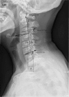
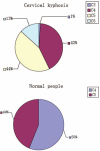
Changes of cervical sagittal alignment during motion in cervical kyphosis patients have never been published before. This study was to investigate the changes and provide a better reference for orthopedic treatment.Randomized double-blind repeat trial was carried out on 60 patients with cervical kyphosis. On standard position, hyper flexion, and hyper extension sagittal radiographs, the following measurements were made: the C2-7 vertebral body spatial alignment angle (∠A), C2-7 vertebral lower terminal lamina tilt angle (∠B), C2/3 to C6/7 segmental intervertebral space angle (∠C), the distance from the posterior edge of odontoid to C7 vertebral body (D value), and the difference of angle A, B, and C between cervical flexion and extension movement. Another 60 healthy volunteers were enrolled, of whom the cervical curve apex was determined using Borden's method to compare change and distribution characteristics to patients with cervical kyphosis and C value.In standard lateral position, ∠A was positive and increased from C2 to C7. In hyper extension position, ∠A decreased with reducing amplitude from C2 to C7 compared with the standard position, whereas in hyper flexion position, the average value of ∠A increased with decreasing amplitude from C2 to C7. ∠B followed similar change regularities as ∠A with a larger mean value. In cervical flexion and extension movement, ∠A change of upper vertebral body (∠D) was almost equal to ∠A change of lower vertebral body and ∠C change between the adjacent 2 vertebral bodies (∠E). The curve apex distribution was almost between C4 and C5 in cervical kyphosis patients. A significant difference was observed between cervical kyphosis patients and normal people in C value and D value.The correction of the cervical kyphosis can be carried out from the apex of the cervical spine that provides a solid theoretical foundation for the correction of the cervical kyphosis.
Copyright © 2017 The Authors. Published by Wolters Kluwer Health, Inc. All rights reserved.
Figures


Similar articles
-
Clinical significance of cervical vertebral flexion and extension spatial alignment changes.Spine (Phila Pa 1976). 2009 Jan 1;34(1):E21-6. doi: 10.1097/BRS.0b013e31818938f5. Spine (Phila Pa 1976). 2009. PMID: 19127144
-
Quantitatively assessing the effect of cervical sagittal alignment on dynamic intervertebral kinematics by video-fluoroscopy technique.Musculoskelet Sci Pract. 2024 Aug;72:102959. doi: 10.1016/j.msksp.2024.102959. Epub 2024 Apr 11. Musculoskelet Sci Pract. 2024. PMID: 38626497
-
Evaluation of sagittal alignment and range of motion of the cervical spine using multi-detector- row computed tomography in asymptomatic subjects.Nagoya J Med Sci. 2018 Nov;80(4):583-589. doi: 10.18999/nagjms.80.4.583. Nagoya J Med Sci. 2018. PMID: 30587872 Free PMC article.
-
Pediatric cervical kyphosis in the MRI era (1984-2008) with long-term follow up: literature review.Childs Nerv Syst. 2022 Feb;38(2):361-377. doi: 10.1007/s00381-021-05409-z. Epub 2021 Nov 22. Childs Nerv Syst. 2022. PMID: 34806157 Review.
-
Cervical sagittal balance: a biomechanical perspective can help clinical practice.Eur Spine J. 2018 Feb;27(Suppl 1):25-38. doi: 10.1007/s00586-017-5367-1. Epub 2017 Nov 6. Eur Spine J. 2018. PMID: 29110218 Review.
Cited by
-
The effect of cervical spine flexion-extension motion on odontoid parameters.J Orthop Surg Res. 2025 Jan 19;20(1):68. doi: 10.1186/s13018-025-05488-7. J Orthop Surg Res. 2025. PMID: 39828697 Free PMC article.
-
Surgical sequence in anterior column realignment with posterior osteotomy is important for degree of adult spinal deformity correction: advantages and indications for posterior to anterior sequence.BMC Musculoskelet Disord. 2022 Nov 22;23(1):1004. doi: 10.1186/s12891-022-05915-4. BMC Musculoskelet Disord. 2022. PMID: 36419151 Free PMC article.
-
Restoring cervical lordosis by cervical extension traction methods in the treatment of cervical spine disorders: a systematic review of controlled trials.J Phys Ther Sci. 2021 Oct;33(10):784-794. doi: 10.1589/jpts.33.784. Epub 2021 Oct 13. J Phys Ther Sci. 2021. PMID: 34658525 Free PMC article. Review.
References
-
- Takeshima T, Omokawa S, Takaoka T, et al. Sagittal alignment of cervical flexion and extension: lateral radiographic analysis. Spine (Phila Pa 1976) 2002;27:E348–355. - PubMed
-
- Boyle JJ, Milne N, Singer KP. Influence of age on cervicothoracic spinal curvature: an ex vivo radiographic survey. Clin Biomech (Bristol, Avon) 2002;17:361–7. - PubMed
-
- <Cobb Method or Harrison Posterior Tangent Method.pdf>. - PubMed
-
- Steinmetz MP, Stewart TJ, Kager CD, et al. Cervical deformity correction. Neurosurgery 2007;60(1 Supp1 1):S90–7. - PubMed
-
- Sharan AD, Krystal JD, Singla A, et al. Advances in the understanding of cervical spine deformity. Instr Course Lect 2015;64:417–26. - PubMed
Publication types
MeSH terms
LinkOut - more resources
Full Text Sources
Other Literature Sources
Medical
Miscellaneous

