

Vascular Ehlers-Danlos syndrome with cryptorchidism, recurrent pneumothorax, and pulmonary capillary hemangiomatosis-like foci: A case report
- PMID: 29381997
- PMCID: PMC5708996
- DOI: 10.1097/MD.0000000000008853
Vascular Ehlers-Danlos syndrome with cryptorchidism, recurrent pneumothorax, and pulmonary capillary hemangiomatosis-like foci: A case report
Abstract
Rationale: Vascular Ehlers-Danlos syndrome (vEDS) is a rare autosomal dominant inherited collagen disorder caused by defects or deficiency of pro-alpha 1 chain of type III procollagen encoded by COL3A1. vEDS is characterized not only by soft tissue manifestations including hyperextensibility of skin and joint hypermobility but also by early mortality due to rupture of arteries or vital organs. Although pulmonary complications are not common, vEDS cases complicated by pneumothorax, hemothorax, or intrapulmonary hematoma have been reported. When a patient initially presents only with pulmonary complications, it is not easy for clinicians to suspect vEDS.
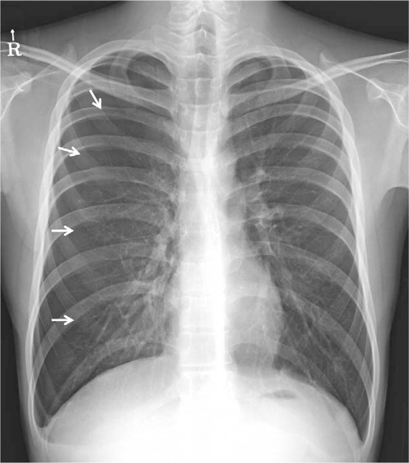
Patient concerns: We report a case of an 18-year-old high school student, with a past history of cryptorchidism, presenting with recurrent pneumothorax.
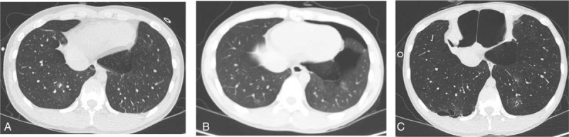
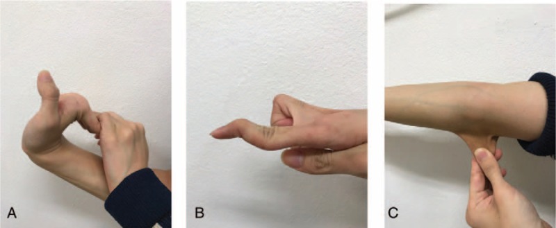
Diagnoses: Routine laboratory findings were unremarkable. Chest high resolution computed tomographic scan showed age-unmatched hyperinflation of both lungs, atypical cystic changes and multifocal ground glass opacities scattered in both lower lobes. His slender body shape, hyperflexible joints, and hyperextensible skin provided clue to suspicion of a possible connective tissue disorder.
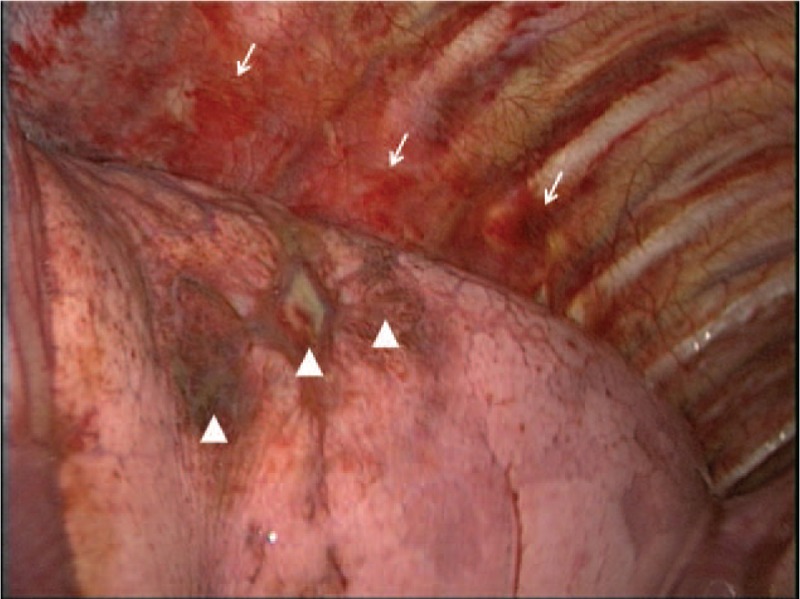
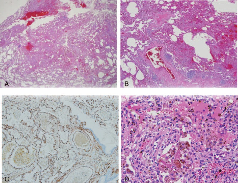
Interventions: The histological examination of the lung lesions showed excessive capillary proliferation in the pulmonary interstitium and pleura allowing the diagnosis of pulmonary capillary hemangiomatosis (PCH)-like foci. Genetic study revealed COL3A1 gene splicing site mutation confirming his diagnosis as vEDS.
Outcomes: Although his diagnosis vEDS is notorious for fatal vascular complication, there was no evidence of such complication at presentation. Fortunately, he has been followed up for 10 months without pulmonary or vascular complications.
Lessons: To the best of our knowledge, both cryptorchidism and PCH-like foci have never been reported yet as complications of vEDS, suggesting our case might be a new variant of this condition. This case emphasizes the importance of comprehensive physical examination and history-taking, and the clinical suspicion of a possible connective tissue disorder when we encounter cases with atypical presentation and/or unique chest radiologic findings especially in young patients.
Copyright © 2017 The Authors. Published by Wolters Kluwer Health, Inc. All rights reserved.
Conflict of interest statement
The authors have no funding and conflicts of interest to disclose.
Figures
Similar articles
-
Recurrent pneumothorax and intrapulmonary cavitary lesions in a male patient with vascular Ehlers-Danlos syndrome and a novel missense mutation in the COL3A1 gene: a case report.BMC Pulm Med. 2020 May 29;20(1):149. doi: 10.1186/s12890-020-1164-4. BMC Pulm Med. 2020. PMID: 32471395 Free PMC article.
-
Spontaneous pneumothorax and hemothorax frequently precede the arterial and intestinal complications of vascular Ehlers-Danlos syndrome.Am J Med Genet A. 2019 May;179(5):797-802. doi: 10.1002/ajmg.a.61094. Epub 2019 Feb 22. Am J Med Genet A. 2019. PMID: 30793832
-
Case report of a young male, with recurrent pneumothorax, hemoptysis and intrapulmonary cavitary lesions.Medicine (Baltimore). 2023 Oct 6;102(40):e35436. doi: 10.1097/MD.0000000000035436. Medicine (Baltimore). 2023. PMID: 37800821 Free PMC article.
-
Vascular Ehlers-Danlos Syndrome With a Novel Missense COL3A1 Mutation Present With Pulmonary Complications and Iliac Arterial Dissection.Vasc Endovascular Surg. 2018 Feb;52(2):138-142. doi: 10.1177/1538574417745432. Epub 2017 Dec 7. Vasc Endovascular Surg. 2018. PMID: 29216800 Review.
-
[Respiratory manifestations of Ehlers-Danlos syndromes].Rev Mal Respir. 2023 Mar;40(3):254-264. doi: 10.1016/j.rmr.2023.01.009. Epub 2023 Feb 4. Rev Mal Respir. 2023. PMID: 36740495 Review. French.
Cited by
-
Pulmonary Mycobacterium avium complex infection with vascular Ehlers-Danlos syndrome: A case report.Respir Med Case Rep. 2024 Sep 16;52:102119. doi: 10.1016/j.rmcr.2024.102119. eCollection 2024. Respir Med Case Rep. 2024. PMID: 39350959 Free PMC article.
-
Urogenital and pelvic complications in the Ehlers-Danlos syndromes and associated hypermobility spectrum disorders: A scoping review.Clin Genet. 2020 Jan;97(1):168-178. doi: 10.1111/cge.13624. Epub 2019 Sep 1. Clin Genet. 2020. PMID: 31420870 Free PMC article.
-
A review of respiratory manifestations and their management in Ehlers-Danlos syndromes and hypermobility spectrum disorders.Chron Respir Dis. 2021 Jan-Dec;18:14799731211025313. doi: 10.1177/14799731211025313. Chron Respir Dis. 2021. PMID: 34291699 Free PMC article. Review.
-
Pulmonary capillary haemangiomatosis: a distinct entity?Eur Respir Rev. 2020 May 27;29(156):190168. doi: 10.1183/16000617.0168-2019. Print 2020 Jun 30. Eur Respir Rev. 2020. PMID: 32461209 Free PMC article.
References
-
- Abel MD, Carrasco LR. Ehlers-Danlos syndrome: classifications, oral manifestations, and dental considerations. Oral Surg Oral Med Oral Pathol Oral Radiol Endod 2006;102:582–90. - PubMed
-
- Beighton P, De Paepe A, Steinmann B, et al. Ehlers-Danlos syndromes: revised nosology, Villefranche, 1997. Ehlers-Danlos National Foundation (USA) and Ehlers-Danlos Support Group (UK). Am J Med Genet 1998;77:31–7. - PubMed
-
- Pepin M, Schwarze U, Superti-Furga A, et al. Clinical and genetic features of Ehlers-Danlos syndrome type IV, the vascular type. N Engl J Med 2000;342:673–80. - PubMed
-
- Dar RA, Wani SH, Mushtaque M, et al. Spontaneous hemo-pneumothorax in a patient with Ehlers-Danlos syndrome. Gen Thorac Cardiovasc Surg 2012;60:587–9. - PubMed
-
- Ishiguro T, Takayanagi N, Kawabata Y, et al. Ehlers-Danlos syndrome with recurrent spontaneous pneumothoraces and cavitary lesion on chest X-ray as the initial complications. Intern Med 2009;48:717–22. - PubMed
Publication types
MeSH terms
Substances
Supplementary concepts
LinkOut - more resources
Full Text Sources
Other Literature Sources
Medical
Miscellaneous
