

Microarchitecture of the Augmented Bone Following Sinus Elevation with an Albumin Impregnated Demineralized Freeze-Dried Bone Allograft (BoneAlbumin) versus Anorganic Bovine Bone Mineral: A Randomized Prospective Clinical, Histomorphometric, and Micro-Computed Tomography Study
- PMID: 29382095
- PMCID: PMC5848899
- DOI: 10.3390/ma11020202
Microarchitecture of the Augmented Bone Following Sinus Elevation with an Albumin Impregnated Demineralized Freeze-Dried Bone Allograft (BoneAlbumin) versus Anorganic Bovine Bone Mineral: A Randomized Prospective Clinical, Histomorphometric, and Micro-Computed Tomography Study
Abstract
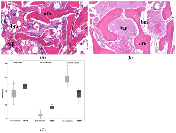
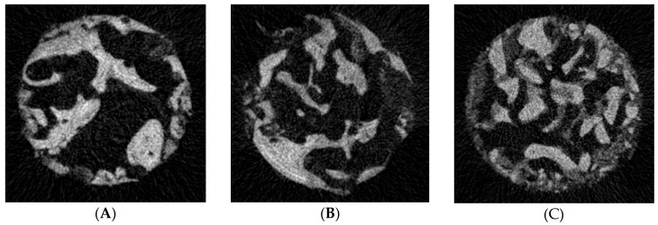
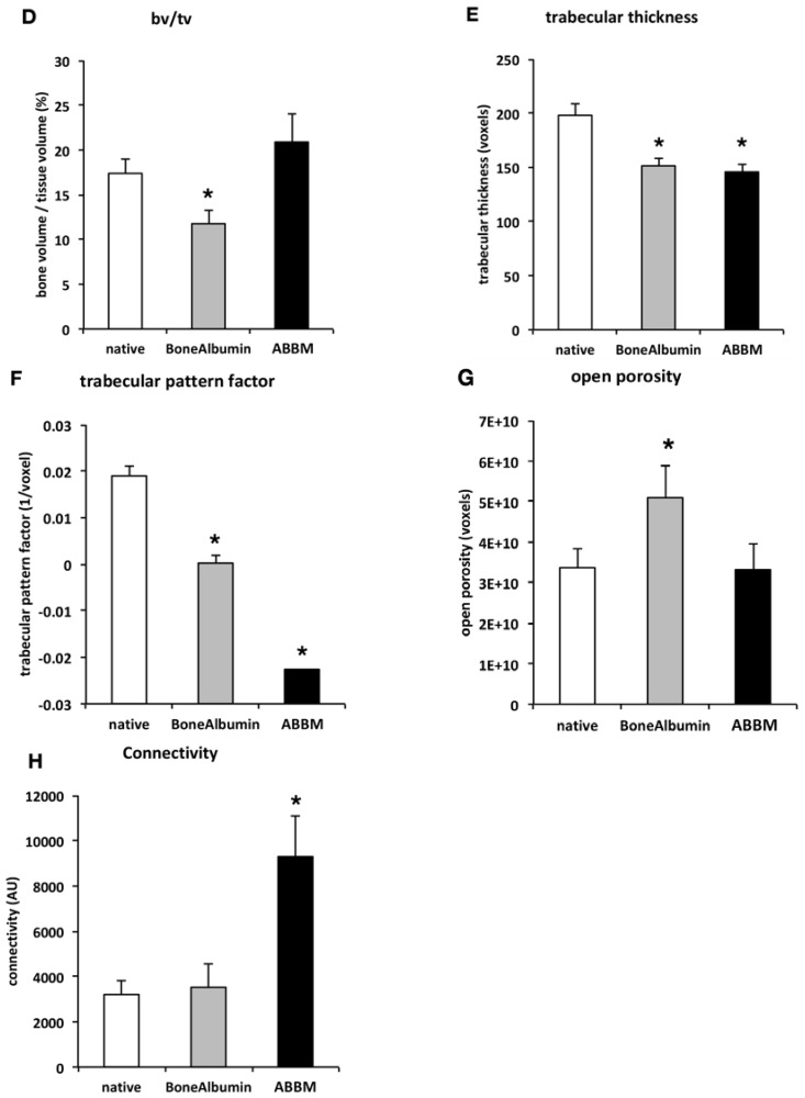
Serum albumin has been identified as an endogenous protein that is integral to early bone regeneration. We hypothesized that albumin addition to allografts may result in better bone remodeling than what can be achieved with anorganic xenografts. Sinus elevations were performed at 32 sites of 18 patients with the lateral window technique. Sites either received filling with an anorganic bovine bone mineral (ABBM, BioOss, Geistlich, CH) or albumin impregnated allograft (BoneAlbumin, OrthoSera, AT). After 6-months patients received dental implants and 16 bone core biopsy samples were obtained from the ABBM filled, and 16 from the BoneAlbumin augmented sites. The biopsies were examined by histomorphometry and µCT. Percentage of the residual graft in the BoneAlbumin group was 0-12.7%, median 5.4% vs. ABBM 6.3-35.9%, median 16.9%, p < 0.05. Results of the µCT analysis showed that the microarchitecture of the augmented bone in the BoneAlbumin group resembles that of the native maxilla in morphometric parameters Trabecular Pattern Factor and Connectivity. Our data show that while ABBM successfully integrates into the newly formed bone tissue as persisting particles, BoneAlbumin is underway towards complete remodeling with new bone closely resembling that of the intact maxilla.
Keywords: allograft; bone graft material; histology; histomorphometry; maxillary sinus floor augmentation; x-ray microtomography; xenograft.
Conflict of interest statement
Author Lacza Zsombo is an inventor on a patent covering albumin coating and holds stock in OrthoSera GmbH, a startup that is commercializing the technology. All other authors declare no conflict of interest. The founding sponsors had no role in the design of the study; in the collection, analyses, or interpretation of data; in the writing of the manuscript, and in the decision to publish the results.
Figures
Similar articles
-
A histomorphometric assessment of collagen-stabilized anorganic bovine bone mineral in maxillary sinus augmentation - a prospective clinical trial.Clin Oral Implants Res. 2016 Jul;27(7):850-8. doi: 10.1111/clr.12694. Epub 2015 Sep 16. Clin Oral Implants Res. 2016. PMID: 26374171 Clinical Trial.
-
Comparison of mineralized cancellous bone allograft (Puros) and anorganic bovine bone matrix (Bio-Oss) for sinus augmentation: histomorphometry at 26 to 32 weeks after grafting.Int J Periodontics Restorative Dent. 2006 Dec;26(6):543-51. Int J Periodontics Restorative Dent. 2006. PMID: 17243327 Clinical Trial.
-
Effect of xenograft (ABBM) particle size on vital bone formation following maxillary sinus augmentation: a multicenter, randomized, controlled, clinical histomorphometric trial.Int J Periodontics Restorative Dent. 2013 Jul-Aug;33(4):467-75. doi: 10.11607/prd.1423. Int J Periodontics Restorative Dent. 2013. PMID: 23820706 Clinical Trial.
-
Histomorphometric Analysis of Maxillary Sinus Grafts: A Pilot Study.Int J Oral Maxillofac Implants. 2019 May/June;34(3):759–767. doi: 10.11607/jomi.6218. Epub 2019 Feb 26. Int J Oral Maxillofac Implants. 2019. PMID: 30807623
-
Clinical evaluation of demineralized bone allograft in a hyaluronic acid carrier for sinus lift augmentation in humans: a computed tomography and histomorphometric study.Clin Oral Implants Res. 2007 Apr;18(2):204-11. doi: 10.1111/j.1600-0501.2006.01303.x. Clin Oral Implants Res. 2007. PMID: 17348885 Clinical Trial.
Cited by
-
A Pilot Clinical Study of Hyperacute Serum Treatment in Osteoarthritic Knee Joint: Cytokine Changes and Clinical Effects.Curr Issues Mol Biol. 2021 Jul 9;43(2):637-649. doi: 10.3390/cimb43020046. Curr Issues Mol Biol. 2021. PMID: 34287260 Free PMC article.
-
Bone Grafting with Albumin-Impregnated Bone Allograft After Odontogenic Cyst Removal.J Clin Med. 2025 Jun 12;14(12):4173. doi: 10.3390/jcm14124173. J Clin Med. 2025. PMID: 40565919 Free PMC article.
-
The Use of Allograft Bone in the Lateral Approach of Sinus Floor Elevation: A Systematic Review of Clinical Studies.Medicina (Kaunas). 2024 Jan 31;60(2):252. doi: 10.3390/medicina60020252. Medicina (Kaunas). 2024. PMID: 38399540 Free PMC article.
-
Comparison between Micro-Computed Tomography and Cone-Beam Computed Tomography in the Assessment of Bone Quality and a Long-Term Volumetric Study of the Augmented Sinus Grafted with an Albumin Impregnated Allograft.J Clin Med. 2020 Jan 21;9(2):303. doi: 10.3390/jcm9020303. J Clin Med. 2020. PMID: 31973237 Free PMC article.
-
Comparison Study of the Histomorphometric Results after Socket Preservation with PRF and Allograft Used for Socket Preservation-Randomized Controlled Trials.Int J Environ Res Public Health. 2021 Jul 13;18(14):7451. doi: 10.3390/ijerph18147451. Int J Environ Res Public Health. 2021. PMID: 34299902 Free PMC article.
References
-
- Kivovics M., Szabo B.T., Nemeth O., Tari N., Dori F., Nagy P., Dobo-Nagy C., Szabo G. Microarchitectural study of the augmented bone following ridge preservation with a porcine xenograft and a collagen membrane: Preliminary report of a prospective clinical, histological, and micro-computed tomography analysis. Int. J. Oral Maxillofac. Surg. 2017;46:250–260. doi: 10.1016/j.ijom.2016.10.010. - DOI - PubMed
LinkOut - more resources
Full Text Sources
Other Literature Sources
