. 2018 Jan 30;20(1):13.
doi: 10.1186/s13075-018-1511-5.

Increase of MZB1 in B cells in systemic lupus erythematosus: proteomic analysis of biopsied lymph nodes

Affiliations

Increase of MZB1 in B cells in systemic lupus erythematosus: proteomic analysis of biopsied lymph nodes

Aya Miyagawa-Hayashino et al. Arthritis Res Ther. .

Abstract

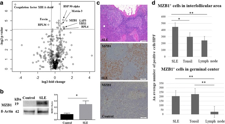
Background: Systemic lupus erythematosus (SLE) is a prototypical autoimmune disease in which dysregulation of B cells has been recognized. Here, we searched for potential biomarkers of SLE using liquid chromatography-tandem mass spectrometry (LC-MS).

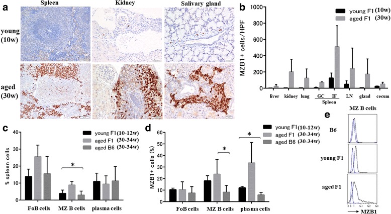
Methods: Lymph nodes from SLE patients and controls were analyzed by LC-MS. To validate the identified molecules, immunoblotting and immunohistochemistry were performed and B cells from SLE patients were analyzed by quantitative RT-PCR. B-cell subsets from NZB/W F1 mice, which exhibit autoimmune disease resembling human SLE, were analyzed by flow cytometry. Endoplasmic reticulum (ER) stress was induced by tunicamycin and the serum concentration of anti-dsDNA antibodies was determined by ELISA. TUNEL methods and immunoblotting were used to assess the effect of tunicamycin.

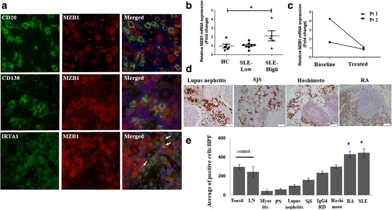
Results: MZB1, which comprises part of a B-cell-specific ER chaperone complex and is a key player in antibody secretion, was one of the differentially expressed proteins identified by LC-MS and confirmed by immunoblotting. Immunohistochemically, larger numbers of MZB1+ cells were located mainly in interfollicular areas and scattered in germinal centers in specimens from SLE patients compared with those from controls. MZB1 colocalized with CD138+ plasma cells and IRTA1+ marginal zone B cells. MZB1 mRNA was increased by 2.1-fold in B cells of SLE patients with active disease (SLE Disease Activity Index 2000 ≥ 6) compared with controls. In aged NZB/W F1 mice, splenic marginal zone B cells and plasma cells showed elevated MZB1 levels. Tunicamycin induced apoptosis of MZB1+ cells in target organs, resulting in decreased serum anti-dsDNA antibody levels. Additionally, MZB1+ cells were increased in synovial tissue specimens from patients with rheumatoid arthritis.

Conclusions: MZB1 may be a potential therapeutic target in excessive antibody-secreting cells in SLE.

Keywords: Formalin-fixed paraffin-embedded; Lupus-prone mice; Proteomic analysis; SLE lymphadenopathy; Systemic lupus erythematosus; TUNEL; Unfolded protein response.


Conflict of interest statement

Authors’ information

Not applicable.

Ethic

Written informed content was obtained from all study participants, and the study was conducted according to the principles expressed in the Declaration of Helsinki. The Ethics Committee of Kyoto University approved this study (Nos. E1872, G520).

Mice were maintained under specific pathogen-free conditions at the Center for Experimental Animals of Kyoto University, and the animal experiments were performed in accordance with the institutional guidelines.

Consent for publication

Not applicable.

Competing interests

TO and YH are employees of Astellas Pharma Inc. This does not alter our adherence to journal policies on sharing data and materials. Astellas Pharma Inc. had no role in the study design or collection, or analysis or interpretation of the data; writing of the manuscript; or the decision to submit the manuscript for publication. Publication of this article was approved by an intellectual property committee composed of representatives from Kyoto University and Astellas Pharma Inc.

Publisher’s Note

Springer Nature remains neutral with regard to jurisdictional claims in published maps and institutional affiliations.

Figures

Fig. 1
Fig. 1
MZB1 is a highly expressed protein in lymph nodes from SLE patients. a Volcano plot showing distribution of all proteins in lymph nodes from SLE patients (n = 3) and controls (n = 3) analyzed by LC-MS. Horizontal line denotes fold change. Vertical line represents p value (ANOVA). Differences > 1.5-fold change and p ≤ 0.05 considered statistically significant. Fold-change values indicate higher (+) and lower (–) expression in SLE patients compared with controls. Significant proteins labeled with their gene name. b Representative immunoblotting for MZB1 in lymph node tissue from SLE patients and controls. Right: Quantification of the immunoblot (n = 3 each). Mean band intensity ratio measured as the intensity of the MZB1 band divided by intensity of the corresponding beta-actin band. Error bars indicate SEM. c Upper: histological section of an axillary lymph node from SLE patients showing reactive follicular hyperplasia. HE, scale bar = 100 μm. Middle: MZB1 immunostaining of the same sample of the upper image, showing numerous positive cells in the interfollicular area and within the germinal center. Scale bar = 50 μm. Lower: MZB1 immunostaining of the control. Scale bar = 50 μm. d MZB1+ cells in lymph nodes from SLE patients were significantly more frequently observed in the germinal center and interfollicular areas compared with those from controls. *p<0.05; **p<0.01 HPF high-power field, SLE systemic lupus erythematosus
Fig. 2
Fig. 2
MZB1 is overexpressed in B-cell subsets and MZB1 mRNA increased in peripheral blood B cells from SLE patients with active disease. a Immunofluorescence showed slight colocalization of MZB1 with B-cell marker CD20 and strong colocalization with plasma cell marker CD138 and MZ B-cell marker IRTA1 in lymph nodes from SLE patients. b MZB1 mRNA levels in peripheral blood B cells from SLE patients with active disease (SLE-High) increased by 2.1-fold compared with those in healthy controls (HC) (p < 0.05). No significant increase in MZB1 mRNA levels observed in peripheral blood B cells from SLE patients with inactive disease (SLE-Low). c Two SLE patients with active disease had follow-up samples collected at 2 months of treatment. Relative MZB1 mRNA expression levels decreased with treatment. d MZB1 immunohistochemistry in tissue from patients with various autoimmune diseases. e Increased proportion of MZB1+ cells observed in lymph nodes from SLE patients and synovial tissue from rheumatoid arthritis (RA) patients compared with control lymph nodes (LN) and tonsils (p < 0.05). RA, scale bar = 50 μm; lupus nephritis, scale bar = 20 μm. Hashimoto Hashimoto’s thyroiditis, HPF high-power field, IgG4-RD IgG4-related pancreatitis, myositis dermatomyositis, PN polyarteritis nodosa, Pt patient, Sjs Sjögren’s syndrome, SLE systemic lupus erythematosus
Fig. 3
Fig. 3
Splenic marginal zone B cells and plasma cells show elevated MZB1 levels in aged lupus-prone mice. a MZB1 immunohistochemistry in young (10 weeks of age) and aged (30 weeks of age) BWF1 mice. Spleen, scale bar = 50 μm; kidney, salivary gland, scale bars = 20 μm. b Increased proportion of MZB1+ cells observed in various organs in aged BWF1 mice compared with young BWF1 mice (ANOVA, p < 0.05). c Spleen cells sorted as B220+CD21high CD23low (MZ B cells) and B220CD138+ (plasma cells). Percentages of B-cell subsets among total spleen cells in young (10–12 weeks of age) and aged (30–34 weeks of age) BWF1 mice compared with those in aged (30–34 weeks of age) B6 mice (n = 3–5 each group). Proportion of total MZ B cells increased on average by 8.8% in aged BWF1 mice compared with young BWF1 and B6 mice (ANOVA, p < 0.05). d MZB1 expression in each B-cell subset in young (10–12 weeks of age) and aged (30–34 weeks of age) BWF1 mice compared with aged (30–34 weeks of age) B6 mice (n = 3–5 each group). MZB1 expression in MZ B cells in aged BWF1 mice significantly higher than that in B6 mice (p < 0.05). In plasma cells, MZB1 expression in aged BWF1 mice significantly higher than that in young BWF1 and B6 mice (ANOVA, p < 0.05). e Representative histogram of MZB1 expression in MZ B cells in aged B6 mice and young and aged BWF1 mice. Blue line represents isotype control. GC germinal center, FoB follicular B, HPF high-power field, gland salivary gland, IF interfollicular area, LN lymph node, MZ B marginal zone B, w weeks, *p<0.05
Fig. 4
Fig. 4
ER stress induces apoptosis of MZB1+ cells in target organs resulting in decreased serum dsDNA. a Baseline expression of MZB1 and BiP assessed on BWF1 mice at ages of 23 and 34 weeks and there were elevated levels at 34 weeks of age. b MZB1 and BiP expression in spleens from BWF1 mice (25 weeks of age) before or after indicated time points (hours and days) following TM treatment. Beta-actin used as a loading control. Representative blot of three independent experiments with similar results. c Number of TUNEL-positive cells per HPF in kidney and salivary gland of aged BWFI mice (30 weeks of age) serially taken after TM treatment was quantified. d (Upper) TUNEL-positive cells present among inflammatory cells in the kidney. (Lower) Condensed and fragmented nuclei in MZB1+ cells from BWF1 mice kidney following TM treatment. e Anti-dsDNA antibody concentrations in BWF1 mice (n = 7; 30 weeks of age) measured serially by ELISA following TM treatment. B6 mice (n = 2; 30 weeks of age) used as control. TUNEL, TdT-mediated dUTP nick end labeling; HPF, high-power field; hr, hour; *p<0.05

References

    1. Tsokos GC. Systemic lupus erythematosus. N Engl J Med. 2011;365:2110–21. doi: 10.1056/NEJMra1100359. - DOI - PubMed
    1. Choi J, Kim ST, Craft J. The pathogenesis of systemic lupus erythematosus—an update. Curr Opin Immunol. 2012;24:651–7. doi: 10.1016/j.coi.2012.10.004. - DOI - PMC - PubMed
    1. Cappione A, 3rd, Anolik JH, Pugh-Bernard A, Barnard J, Dutcher P, Silverman G, Sanz I. Germinal center exclusion of autoreactive B cells is defective in human systemic lupus erythematosus. J Clin Invest. 2005;115:3205–16. doi: 10.1172/JCI24179. - DOI - PMC - PubMed
    1. Merrill JT, Neuwelt CM, Wallace DJ, Shanahan JC, Latinis KM, Oates JC, Utset TO, Gordon C, Isenberg DA, Hsieh HJ, Zhang D, Brunetta PG. Efficacy and safety of rituximab in moderately-to-severely active systemic lupus erythematosus: the randomized, double-blind, phase II/III systemic lupus erythematosus evaluation of rituximab trial. Arthritis Rheum. 2010;62:222–33. doi: 10.1002/art.27233. - DOI - PMC - PubMed
    1. Rovin BH, Furie R, Latinis K, Looney RJ, Fervenza FC, Sanchez-Guerrero J, Maciuca R, Zhang D, Garg JP, Brunetta P, Appel G. Efficacy and safety of rituximab in patients with active proliferative lupus nephritis: the Lupus Nephritis Assessment with Rituximab study. Arthritis Rheum. 2012;64:1215–26. doi: 10.1002/art.34359. - DOI - PubMed

Publication types

MeSH terms