. 2018 Jan 30;6(1):10.
doi: 10.1186/s40425-017-0313-7.

Mathematical modeling of tumor-associated macrophage interactions with the cancer microenvironment

Affiliations

Mathematical modeling of tumor-associated macrophage interactions with the cancer microenvironment

Grace Mahlbacher et al. J Immunother Cancer. .

Abstract

Background: Immuno-oncotherapy has emerged as a promising means to target cancer. In particular, therapeutic manipulation of tumor-associated macrophages holds promise due to their various and sometimes opposing roles in tumor progression. It is established that M1-type macrophages suppress tumor progression while M2-types support it. Recently, Tie2-expressing macrophages (TEM) have been identified as a distinct sub-population influencing tumor angiogenesis and vascular remodeling as well as monocyte differentiation.

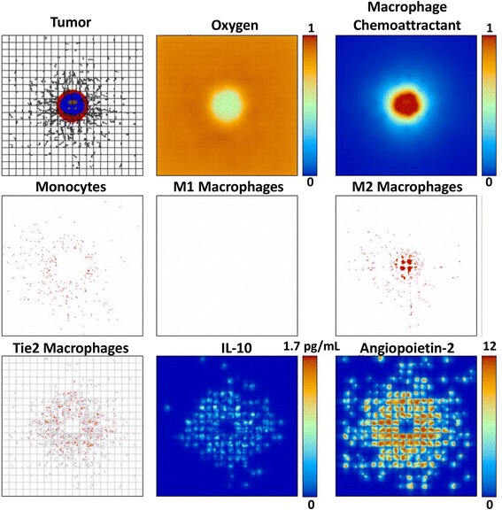
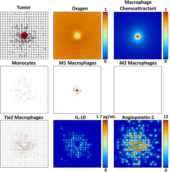
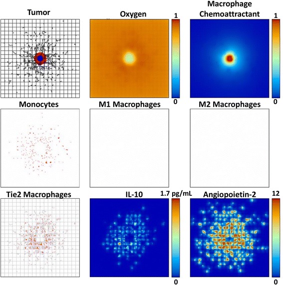
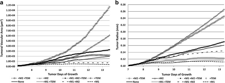
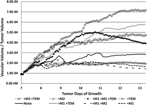
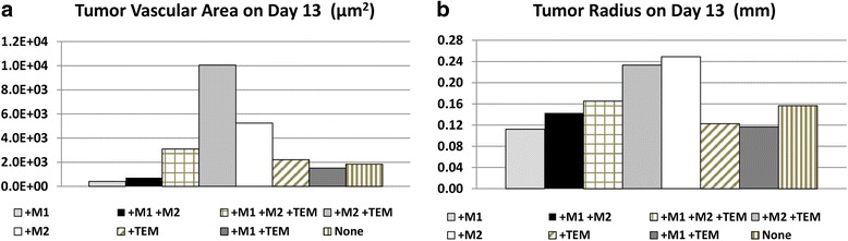
Methods: This study develops a modeling framework to evaluate macrophage interactions with the tumor microenvironment, enabling assessment of how these interactions may affect tumor progression. M1, M2, and Tie2 expressing variants are integrated into a model of tumor growth representing a metastatic lesion in a highly vascularized organ, such as the liver. Behaviors simulated include M1 release of nitric oxide (NO), M2 release of growth-promoting factors, and TEM facilitation of angiogenesis via Angiopoietin-2 and promotion of monocyte differentiation into M2 via IL-10.

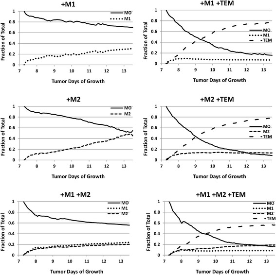
Results: The results show that M2 presence leads to larger tumor growth regardless of TEM effects, implying that immunotherapeutic strategies that lead to TEM ablation may fail to restrain growth when the M2 represents a sizeable population. As TEM pro-tumor effects are less pronounced and on a longer time scale than M1-driven tumor inhibition, a more nuanced approach to influence monocyte differentiation taking into account the tumor state (e.g., under chemotherapy) may be desirable.

Conclusions: The results highlight the dynamic interaction of macrophages within a growing tumor, and, further, establish the initial feasibility of a mathematical framework that could longer term help to optimize cancer immunotherapy.

Keywords: Cancer immunotherapy; Cancer metastasis; Computational simulation; Mathematical modeling; Tie2 expressing macrophages; Tumor-associated macrophages.


Conflict of interest statement

Ethics approval and consent to participate

Not applicable

Consent for publication

Not applicable

Competing interests

The authors declare that they have no competing interests.

Publisher’s Note

Springer Nature remains neutral with regard to jurisdictional claims in published maps and institutional affiliations.

Figures

Fig. 1
Fig. 1
Tumor growth by 13 days with TEM and M1 and M2 macrophage subtypes present. Top left: Tumor with proliferating regions in red and quiescent in blue. Vessels here are shown as black lines, with preexisting vessels comprising the grid space and angiogenic vessels depicted as irregular offshoots. Top center: Oxygen gradient, showing hypoxic conditions in the tumor interior. Top right: Macrophage chemoattractant, (e.g., tumor angiogenesis factors) secreted by the hypoxic tissue of the tumor. Middle left: Monocytes extravasated from the vasculature. Middle center: M1 macrophages shown in red. Middle right: M2 macrophages shown in red. Bottom l eft: TEMs shown in red with vasculature superimposed in gray. The Angiopoietin-2 secreted by the vasculature has caused the TEMs to cluster around the neoangiogenic sprouts. Bottom center: IL-10 secreted by the TEM, which favors monocyte differentiation into M2. Bottom right: Angiopoietin-2 secreted by the neovasculature, which attracts the TEMs to accumulate by the vessels. Each panel represents 4 mm2
Fig. 2
Fig. 2
Tumor growth by 13 days with both TEM and M2 macrophage subtypes present (same description of panels as in Fig. 1). The M2 macrophages penetrate into the tumor following the gradient of macrophage chemoattractants, with their distribution more scattered than the M1 in the vicinity of the tumor due to the monocyte contact with the TEM-eluted IL-10. The tumor is substantially larger than in Fig. 1, and has more hypoxia
Fig. 3
Fig. 3
Tumor growth by 13 days with both TEM and M1 macrophage subtypes present (same description of panels as in Fig. 1). The M1 macrophages penetrate into the tumor following the gradient of macrophage chemoattractants while the TEM remain close to the neovascular network. The tumor is significantly smaller than in Fig. 1, and has less hypoxia
Fig. 4
Fig. 4
Tumor growth by 13 days with only the TEM subtype present (same description of panels as in Fig. 1). In this case, the size of the tumor and its associated hypoxia lie in between the TEM/M2 and TEM/M1 cases
Fig. 5
Fig. 5
Time evolution of the macrophage sub-populations depending on the type of the sub-populations. Left column (from top to bottom): M1-only, M2-only, and M1/M2 cases. Right column: as in left column plus TEM. MO: monocytes
Fig. 6
Fig. 6
Time evolution of the tumor vasculature and tissue as a function of macrophage sub-populations. a Tumor vascular surface area (μm2) calculated using actual capillary radii values influenced by pressure and TEM effects. b Tumor radius (mm). None: no macrophages present
Fig. 7
Fig. 7
Size of tumor vasculature and tumor on day 13 post inception as a function of macrophage sub-populations. a Tumor vascular surface area (μm2) assuming capillary sizes of 10 μm diameter. b Tumor radius (mm). None: no macrophages present
Fig. 8
Fig. 8
Time evolution of ratio of vascular volume to tumor volume as a function of macrophage sub-populations present. None: no macrophages present

References

    1. Chanmee T, Ontong P, Konno K, Itano N. Tumor-associated macrophages as major players in the tumor microenvironment. Cancers. 2014;6(3):1670–1690. doi: 10.3390/cancers6031670. - DOI - PMC - PubMed
    1. De Palma M, Lewis CE. Macrophage regulation of tumor responses to anticancer therapies. Cancer Cell. 2013;23(3):277–286. doi: 10.1016/j.ccr.2013.02.013. - DOI - PubMed
    1. Squadrito ML, De Palma M. Macrophage regulation of tumor angiogenesis: implications for cancer therapy. Mol Asp Med. 2011;32(2):123–145. doi: 10.1016/j.mam.2011.04.005. - DOI - PubMed
    1. Laoui D, Movahedi K, Van Overmeire E, Van den Bossche J, Schouppe E, Mommer C, Nikolaou A, Morias Y, De Baetselier P, Van Ginderachter JA. Tumor-associated macrophages in breast cancer: distinct subsets, distinct functions. The International journal of developmental biology. 2011;55(7–9):861–867. doi: 10.1387/ijdb.113371dl. - DOI - PubMed
    1. Italiani P, Boraschi D. From Monocytes to M1/M2 macrophages: Phenotypical vs. Funct Differ Front Immunol. 2014;5:514. - PMC - PubMed

Publication types

LinkOut - more resources