. 2018 Apr 2;217(4):1369-1382.
doi: 10.1083/jcb.201708044. Epub 2018 Jan 30.

Tom70 enhances mitochondrial preprotein import efficiency by binding to internal targeting sequences

Affiliations

Tom70 enhances mitochondrial preprotein import efficiency by binding to internal targeting sequences

Sandra Backes et al. J Cell Biol. .

Abstract

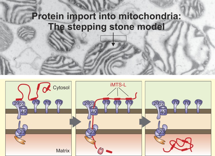
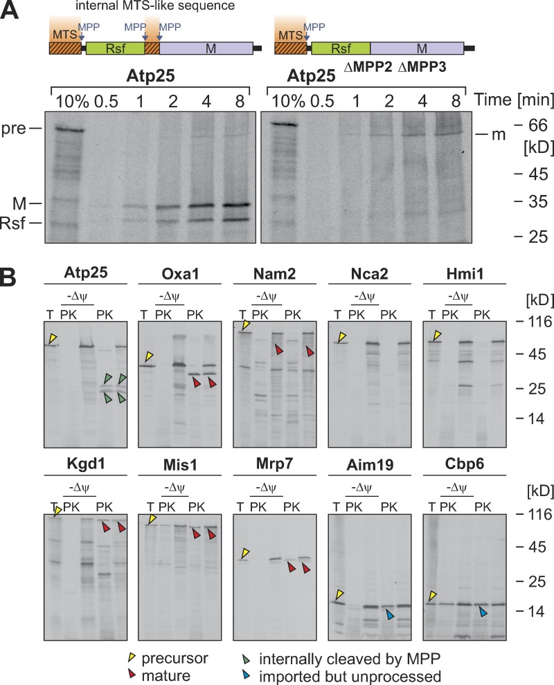
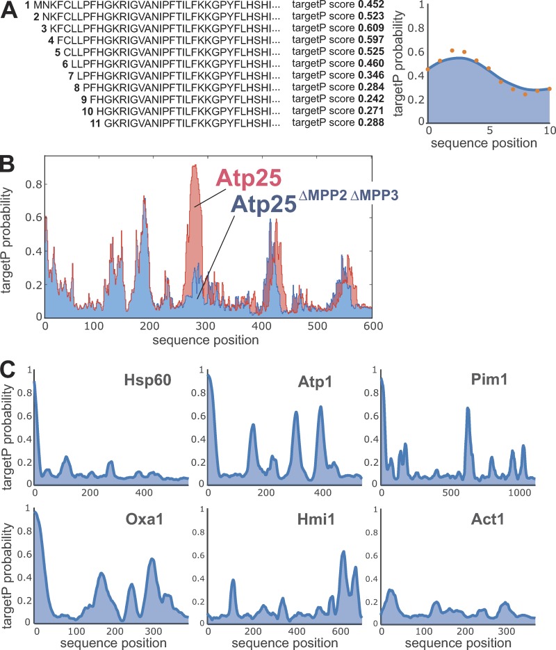
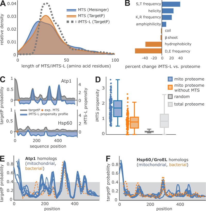
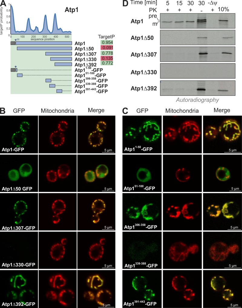
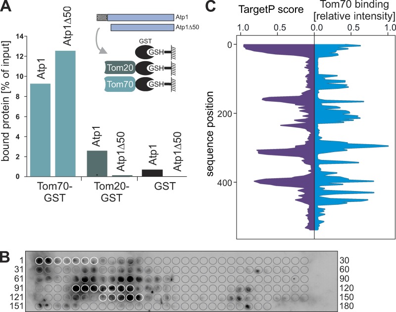
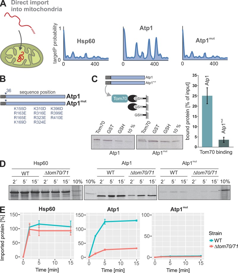
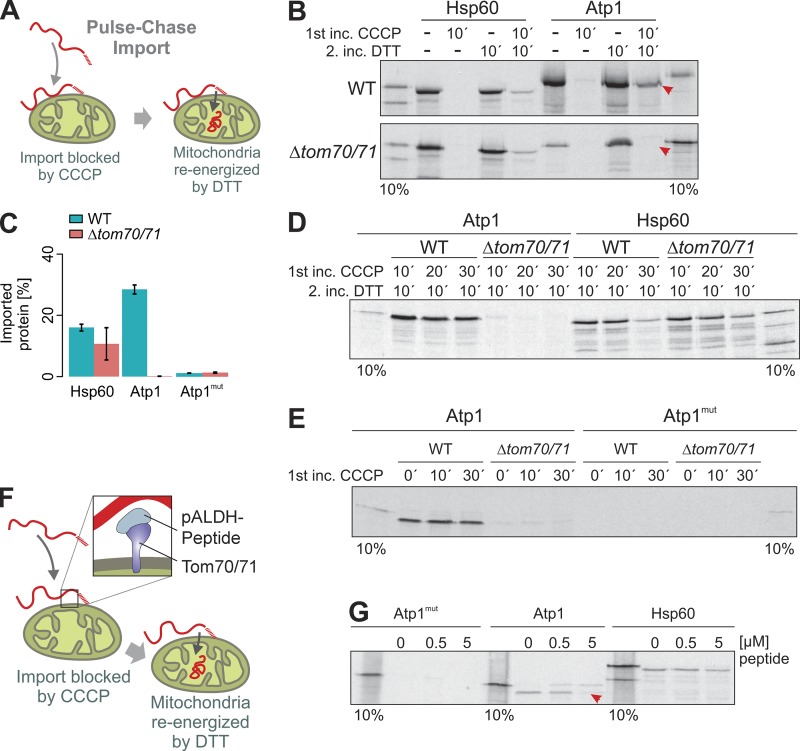
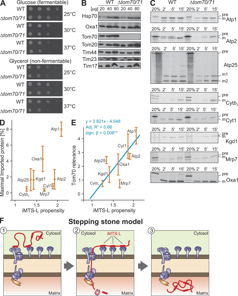
The biogenesis of mitochondria depends on the import of hundreds of preproteins. N-terminal matrix-targeting signals (MTSs) direct preproteins to the surface receptors Tom20, Tom22, and Tom70. In this study, we show that many preproteins contain additional internal MTS-like signals (iMTS-Ls) in their mature region that share the characteristic properties of presequences. These features allow the in silico prediction of iMTS-Ls. Using Atp1 as model substrate, we show that iMTS-Ls mediate the binding to Tom70 and have the potential to target the protein to mitochondria if they are presented at its N terminus. The import of preproteins with high iMTS-L content is significantly impaired in the absence of Tom70, whereas preproteins with low iMTS-L scores are less dependent on Tom70. We propose a stepping stone model according to which the Tom70-mediated interaction with internal binding sites improves the import competence of preproteins and increases the efficiency of their translocation into the mitochondrial matrix.


Figures

None
Graphical abstract
Figure 1.
Figure 1.
Atp25 is a rare example for a tandem precursor protein in yeast. (A) Atp25 and Atp25ΔMPP2ΔMPP3 (lacking amino acids 279–293; Woellhaf et al., 2016) were synthesized in the presence of [35S]methionine and incubated with isolated mitochondria for the times indicated. Nonimported material was removed by degradation with proteinase K (PK) before samples were analyzed by SDS-PAGE and autoradiography. Precursor (pre) and the M and Rsf domains as well as the N-terminally cleaved mature (m) species are indicated. 10% of the radiolabeled precursor protein used per time point was loaded for control. (B) The indicated proteins were radiolabeled and incubated with isolated mitochondria in the presence or absence of membrane potential (Δψ). The samples were split, and one fraction was treated with proteinase K. A 20% total (T) of the precursor protein used per import reaction was loaded for control. Yellow arrowheads depict precursor proteins, red arrowheads indicate N-terminally matured proteins, and green arrowheads indicate internally matured proteins. The presequences of Aim19 and Cbp6 were not cleaved by MPP (Vögtle et al., 2009); the imported species of these proteins are indicated by blue arrowheads. No protease-protected forms of Nca2 and Hmi1 were observed, indicating that these proteins were not imported in our in vitro import system.
Figure 2.
Figure 2.
The TargetP algorithm can be used to predict iMTS-Ls throughout protein sequences. (A) The TargetP algorithm is designed to calculate prediction scores for N-terminal sequences. To calculate scores for internal regions, we consecutively N-terminally truncated the sequences and calculated the corresponding TargetP scores for each position (orange dots), leading to a characteristic profile that shows internal regions in proteins with presequence-like properties. Profiles are smoothed using a Savitzky-Golay filter (blue line). (B) Raw TargetP profiles in Atp25 predict with accuracy the iMTS of the protein, which contains the two internal MPP cleavage sites (Woellhaf et al., 2016). In the Atp25ΔMPP2ΔMPP3 variant, the internal presequence-like region was deleted, and hence, the internal region with the very high TargetP scores is missing. (C) Smoothed TargetP profiles of mitochondrial preproteins without (Hsp60) and with (Atp1, Pim1, Oxa1, and Hmi1) iMTS-Ls. The TargetP profile of the cytosolic protein actin (Act1) is shown for comparison.
Figure 3.
Figure 3.
Many precursor proteins contain iMTS-Ls. (A) Kernel density estimation of the length distribution shows the agreement of experimentally verified MTS length (blue; Vögtle et al., 2009), TargetP-predicted MTS (orange), and the length of iMTS-Ls predicted by our approach (dotted gray line). (B) Sequence property differences between iMTS-Ls and the total yeast proteome are shown in percent change. (C) The iMTS-L propensity profile (blue) for two selected proteins, Atp1 and Hsp60, is plotted against the smoothed TargetP profile (gray). The experimentally verified end of the MTS (Vögtle et al., 2009) is marked (orange dot) and matches the x axis intersection of the corresponding iMTS-L propensity profile. (D) The iMTS-L propensity calculated for experimentally verified mitochondrial matrix proteins (blue; Vögtle et al., 2009) shows a clear difference from random iMTS-L propensity calculated based on a sequence biased by yeast proteome amino acid frequency. However, the iMTS-L propensity of the total yeast proteome is similar to that of mature regions of matrix proteins. (E and F) Atp1 and Hsp60/GroEL sequences of S. cerevisiae (NP_009453 and NP_013360), Kluyveromyces lactis (XP_454248 and XP_455510), Candida glabrata (XP_449761 and XP_448482), S. pombe (CAB11207 and NP_592894), Drosophila (NP_726243 and NP_511115), Homo sapiens (AAH11384 and P10809), Arabidopsis thaliana (P92549 and AEE76842), Chlamydomonas reinhardtii (EDP07337 and XP_001691353), E. coli (WP_021537164 and ABF67773), and Rickettsia prowazekii (WP_004599658 and AMS12509) were aligned. iMTS-L propensity scores of mitochondrial and bacterial homologues were calculated and are shown as blue and orange traces, respectively.
Figure 4.
Figure 4.
iMTS-Ls have mitochondrial targeting potential if present at the N terminus. (A) TargetP profile of Atp1. Overview and TargetP scores of N-terminally truncated Atp1 variants. (B and C) Fusion proteins of N-terminally truncated Atp1 variants and GFP were expressed in yeast cells. Representative fluorescence microscopy images of yeast cells show the intracellular distribution of GFP (green) and mitochondria stained with rhodamine B hexylester (red). Please note that Atp1-GFP, Atp1Δ307-GFP, and Atp1Δ392-GFP as well as Atp11–50-GFP, Atp1306–358-GFP, and Atp1391–443-GFP colocalize with mitochondria, whereas Atp1Δ50-GFP and Atp11–50-GFP are present in the cytosol. Atp1Δ330-GFP and Atp1339–388-GFP failed to be expressed. (D) N-terminally truncated variants of Atp1 were synthesized in reticulocyte lysate in the presence of [35S]methionine and incubated for different times with isolated WT mitochondria. For the sample labeled with –ψ, the mitochondrial membrane potential was dissipated by the addition of valinomycin. Nonimported protein was removed by treatment with proteinase K (PK) before the samples were visualized by SDS-PAGE and autoradiography. 10% of the radiolabeled protein used per import lane was shown for control. Atp1 was efficiently imported into mitochondria. Only very minor amounts of Atp1Δ307-GFP and Atp1Δ392-GFP were taken up, and Atp1Δ50-GFP was not imported. Atp1Δ330-GFP failed to be synthesized.
Figure 5.
Figure 5.
Tom70 binds to specific regions in the mature part of Atp1. (A) The cytosolic receptor domains of Tom70 and Tom20 were purified as GST fusion proteins and bound to GSH beads. Radiolabeled Atp1 or Atp1Δ50 was incubated with the indicated beads for 10 min at room temperature. Beads were then pelleted by centrifugation, the supernatant fractions were discarded, and the pellet fractions were analyzed by SDS-PAGE and autoradiography. The results were quantified using ImageJ. (B) 20-mer peptides shifted by three amino acids along the entire 545 residues of the Atp1 sequence were covalently bound to a membrane and incubated with purified Tom70-GST as used in A. After extensive washing, the bound Tom70 was detected by Western blotting using a specific GST antibody. Circles indicate the location of each single peptide on the membrane. White circles represent the peptides of the N-terminal presequence of Atp1 (peptides 1–7) and the iMTS-L sequences presented in Fig. 4 A (peptides 95–101 and 128–132). (C) Binding of Tom70-GST was quantified, and the relative intensity of each peptide spot was plotted (blue trace). For comparison, the profile of Atp1 TargetP scores is shown (purple trace).
Figure 6.
Figure 6.
The presence of MTS-like sequences in the mature part of mitochondrial proteins influences the import behavior. (A and B) TargetP probabilities of Hsp60, Atp1, and an Atp1mut version of Atp1 in which the iMTS-Ls were mutated by replacing positively charged residues by negative ones. (C) Radiolabeled Atp1 or Atp1mut precursor was incubated with empty GSH beads or beads coupled with GST or GST-Tom70. Beads were pelleted by centrifugation, extensively washed, and analyzed by SDS-PAGE and autoradiography. Lanes labeled “10%” show 10% of the radiolabeled precursor proteins used per reaction. The levels of proteins bound to Tom70 relative to those bound to empty beads were quantified from three independent experiments. Shown are means and SD. (D) The indicated model proteins were radiolabeled in reticulocyte lysate and incubated for the indicated times with WT and Δtom70/Δtom71 mitochondria at 25°C. Mitochondria were incubated with 100 µg/ml proteinase K for 30 min on ice to remove nonimported material and analyzed by SDS-PAGE and autoradiography. Lanes labeled “10%” show 10% of the radiolabeled precursor proteins used per time point. (E) The experiments shown in C were repeated three times and quantified. Intensities were normalized to the 10% control of the corresponding precursor protein. Means and SEM are shown.
Figure 7.
Figure 7.
Tom70 is essential to maintain the Atp1 precursor in an import-competent conformation. (A) Model of the pulse-chase assay used in the following experiments. The membrane potential of isolated mitochondria was dissipated by addition of CCCP. Radiolabeled precursor proteins were added. After incubation of the mitochondria for 10–30 min, CCCP was quenched by DTT, and the mitochondria were reenergized. (B–E) WT or Δtom70/71 mitochondria were preincubated with CCCP for 10–30 min in the presence of radiolabeled Hsp60, Atp1, and Atp1mut (pulse). Mitochondria were reisolated and reenergized by treatment with DTT. After incubation (chase) for 10 min at 25°C, nonimported protein was removed by protease treatment, and samples were analyzed by SDS-PAGE and autoradiography. C shows means and SEM of three replicates. Arrowheads indicate the Atp1 protein that only was chased into mitochondria if Tom70/Tom71 were present. (F and G) A peptide from the rat pALDH presequence that was shown to work as a competitive inhibitor of the Tom70 receptor (Melin et al., 2015) was added to isolated mitochondria to the pulse-chase reaction with Atp1mut, Atp1, and Hsp60 precursors. The presence of 5 µM pALDH peptide prevented the import of Atp1 (red arrowheads).
Figure 8.
Figure 8.
The presence of Tom70 supports the import of precursor proteins with high iMTS-L propensity scores. (A) WT and Δtom70/Δtom71 were grown to log phase in galactose medium before tenfold serial dilutions were spotted onto plates containing glucose or glycerol medium. Plates were incubated at the indicated temperatures for 2 d. Mutants lacking Tom70 and its paralog Tom71 show growth defects at 37°C. (B) Mitochondria were isolated from WT and Δtom70/Δtom71 cells. 20, 40, and 80 µg of mitochondrial proteins were resolved by SDS-PAGE and analyzed by Western blotting. (C) The indicated proteins were radiolabeled in reticulocyte lysate and incubated for the indicated times with WT and Δtom70/Δtom71 mitochondria at 25°C. Mitochondria were reisolated, incubated with 100 µg/ml proteinase K for 30 min on ice to remove nonimported material, and analyzed by SDS-PAGE and autoradiography. Lanes labeled “20%” show 20% of the radiolabeled precursor proteins used per time point. (D) The import experiments shown in C were repeated three times. The signals were quantified and fitted with the Michaelis-Menten equation y = (x + imax)/(x + i50%) from which maximal imported amounts imax were deduced (Fig. S2). Absolute amounts of imported proteins and iMTS-L propensities show no significant relatedness (y = 5.051x − 5.399; adjusted R2 = 0.23; signal β = 0.125). (E) In contrast, regression analysis revealed a significant relationship between the iMTS-L propensity of a protein and its Tom70 relevance (y = 3.821x − 4.048; adjusted R2 = 0.66; signal β = 0.008**), which we define as the protein maximally imported into WT mitochondria divided by that of Δtom70tom71 mitochondria. Error bars denote SD. (F) Hypothetical model for the role of iMTS-Ls as stepping stones for Tom70 interaction during protein import. Binding of Tom70 to the iMTS-Ls prevents premature folding or aggregation of the precursors and maintains their import competence.

References

    1. Alikhani, N., Berglund A.K., Engmann T., Spånning E., Vögtle F.N., Pavlov P., Meisinger C., Langer T., and Glaser E.. 2011. Targeting capacity and conservation of PreP homologues localization in mitochondria of different species. J. Mol. Biol. 410:400–410. 10.1016/j.jmb.2011.05.009 - DOI - PubMed
    1. Altmann, K., Dürr M., and Westermann B.. 2007. Saccharomyces cerevisiae as a model organism to study mitochondrial biology. In Mitochondria. Practical Protocols. Vol. 372. Leister D., and Herrmann J.M., editors. Humana Press, Totowa, New Jersey. 81–90. 10.1007/978-1-59745-365-3_6 - DOI - PubMed
    1. Backes, S., and Herrmann J.M.. 2017. Protein Translocation into the Intermembrane Space and Matrix of Mitochondria: Mechanisms and Driving Forces. Front. Mol. Biosci. 4:83. 10.3389/fmolb.2017.00083 - DOI - PMC - PubMed
    1. Baker, A., and Schatz G.. 1987. Sequences from a prokaryotic genome or the mouse dihydrofolate reductase gene can restore the import of a truncated precursor protein into yeast mitochondria. Proc. Natl. Acad. Sci. USA. 84:3117–3121. 10.1073/pnas.84.10.3117 - DOI - PMC - PubMed
    1. Banerjee, R., Gladkova C., Mapa K., Witte G., and Mokranjac D.. 2015. Protein translocation channel of mitochondrial inner membrane and matrix-exposed import motor communicate via two-domain coupling protein. eLife. 4:e11897. 10.7554/eLife.11897 - DOI - PMC - PubMed

Publication types

MeSH terms

Substances