. 2018 Mar 23;293(12):4532-4544.
doi: 10.1074/jbc.RA117.000732. Epub 2018 Jan 30.

Dimerization of sortilin regulates its trafficking to extracellular vesicles

Affiliations

Dimerization of sortilin regulates its trafficking to extracellular vesicles

Shinsuke Itoh et al. J Biol Chem. .

Abstract

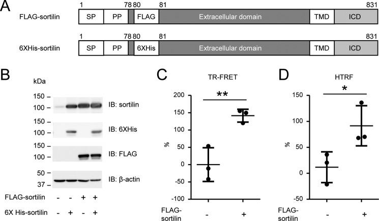
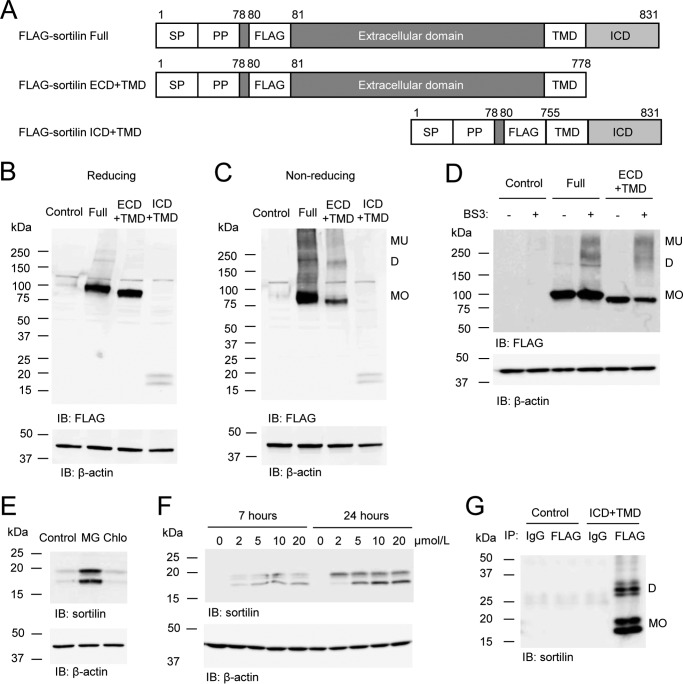
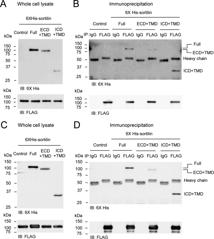
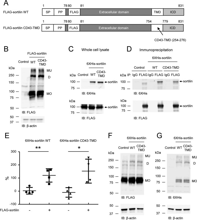
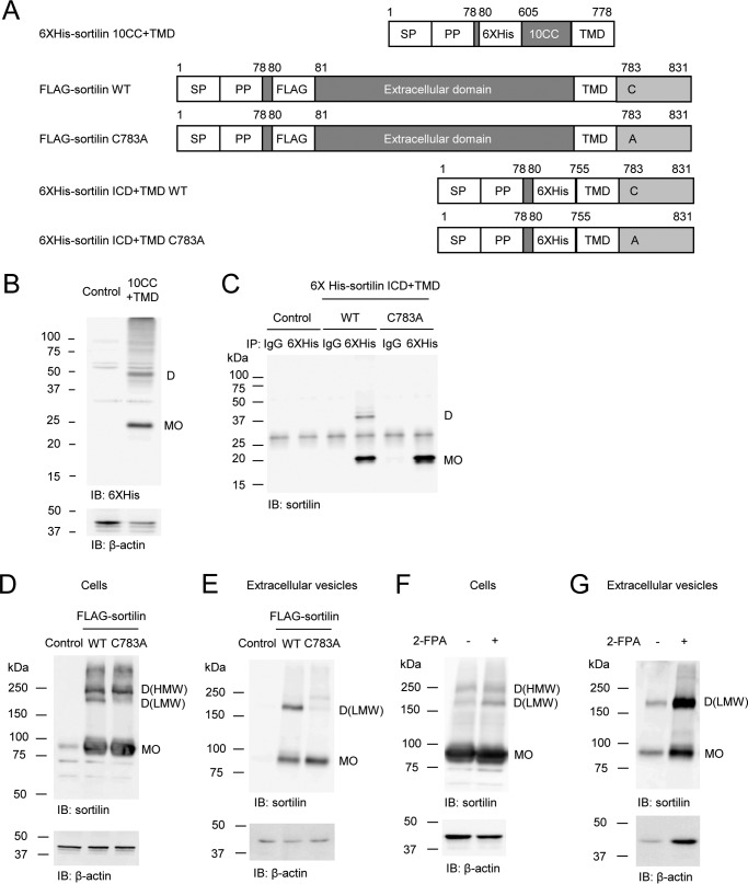
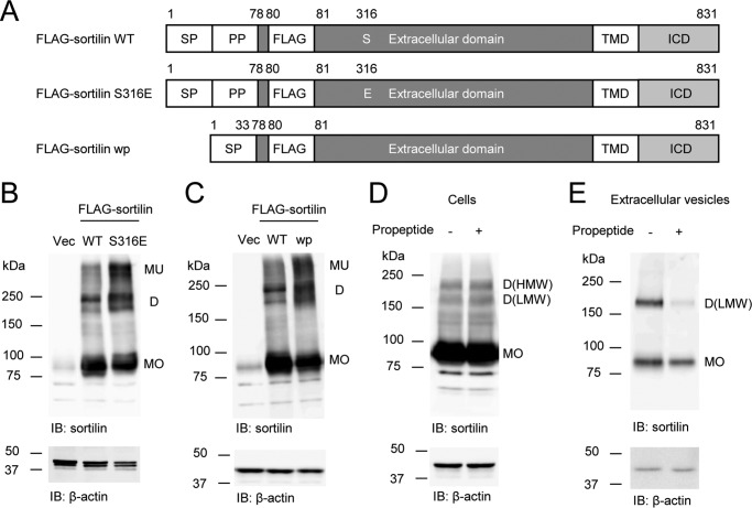
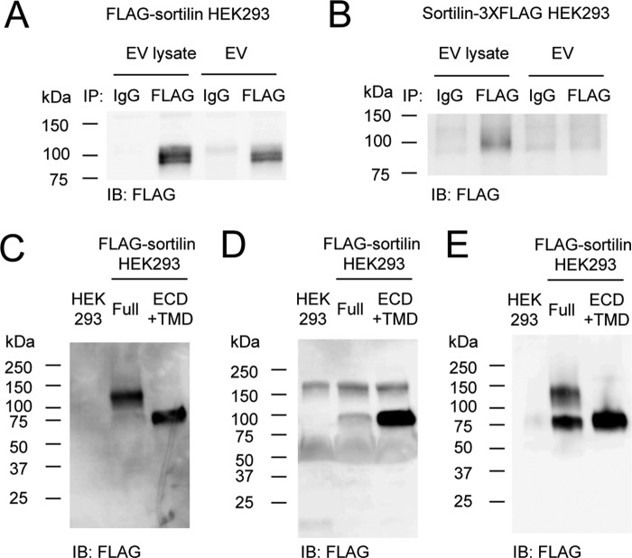
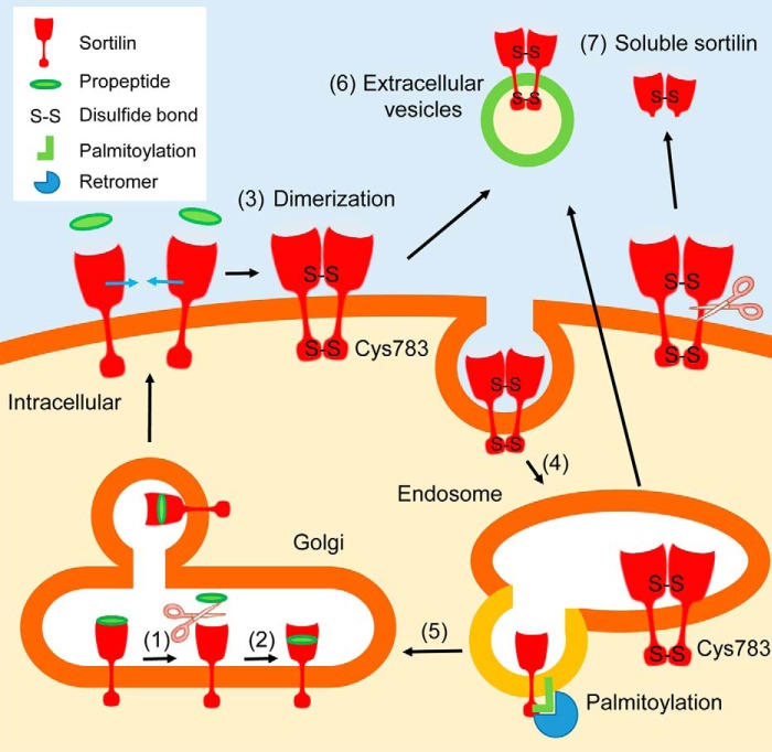
Extracellular vesicles (EVs) play a critical role in intercellular communication by transferring microRNAs, lipids, and proteins to neighboring cells. Sortilin, a sorting receptor that directs target proteins to the secretory or endocytic compartments of cells, is found in both EVs and cells. In many human diseases, including cancer and cardiovascular disorders, sortilin expression levels are atypically high. To elucidate the relationship between cardiovascular disease, particularly vascular calcification, and sortilin expression levels, we explored the trafficking of sortilin in both the intracellular and extracellular milieu. We previously demonstrated that sortilin promotes vascular calcification via its trafficking of tissue-nonspecific alkaline phosphatase to EVs. Although recent reports have noted that sortilin is regulated by multiple post-translational modifications, the precise mechanisms of sortilin trafficking still need to be determined. Here, we show that sortilin forms homodimers with an intermolecular disulfide bond at the cysteine 783 (Cys783) residue, and because Cys783 can be palmitoylated, it could be shared via palmitoylation and an intermolecular disulfide bond. Formation of this intermolecular disulfide bond leads to trafficking of sortilin to EVs by preventing palmitoylation, which further promotes sortilin trafficking to the Golgi apparatus. Moreover, we found that sortilin-derived propeptide decreased sortilin homodimers within EVs. In conclusion, sortilin is transported to EVs via the formation of homodimers with an intermolecular disulfide bond, which is endogenously regulated by its own propeptide. Therefore, we propose that inhibiting dimerization of sortilin acts as a new therapeutic strategy for the treatment of EV-associated diseases, including vascular calcification and cancer.

Keywords: calcification; dimerization; extracellular vesicles; intermolecular disulfide bond; sortilin; sorting; trafficking.


Conflict of interest statement

S. I. and K. M. are employees of Kowa Company, Ltd

Figures

Figure 1.
Figure 1.
Sortilin forms homodimers on the cell surface of HEK293 cells. A, schematic of FLAG-sortilin and His6-sortilin. FLAG tag and His6 tag were inserted following propeptide and 3 amino acids (Ser78-Ala79-Pro80) in sortilin. SP, signal peptide; PP, propeptide. B, overexpression of FLAG-sortilin and His6-sortilin in HEK293 cells was validated by Western blotting. C and D, detection of binding of FLAG-sortilin and His6-sortilin on the cell surface of HEK293 in TR-FRET assay (C) and HTRF assay (D). Change of FRET signal by expression of His6-sortilin is indicated by percent change (mean ± S.D., three independent experiments). Error bars represent S.D. *, p < 0.05; **, p < 0.01 by t test. IB, immunoblotting.
Figure 2.
Figure 2.
Sortilin forms homodimers in the extracellular and intracellular domains with intermolecular disulfide bonds in HEK293 cells. A, schematic of FLAG-sortilin Full, ECD+TMD, and ICD+TMD. SP, signal peptide; PP, propeptide. B and C, protein expression of FLAG-sortilin Full, ECD+TMD, and ICD+TMD was validated in reducing (B) and non-reducing (C) Western blotting using anti-FLAG antibody. FLAG-sortilin Full and ECD+TMD form homodimers and multimers. Empty vector was used as a control. D, HEK293 cells transiently overexpressing FLAG-sortilin Full or ECD+TMD were treated with a cross-linker, BS3, and the cell lysates were used for reducing Western blotting with anti-FLAG antibody, showing dimerization of sortilin Full and ECD+TMD (n = 3). E, HEK293 cells stably overexpressing FLAG-sortilin ICD+TMD (FLAG-sortilin ICD+TMD HEK293 cells) were incubated with DMSO (Control), 20 μmol/liter MG-132 (MG) or 10 μmol/liter chloroquine (Chlo) for 7 h, and then reducing Western blotting was performed using anti-sortilin antibody. MG-132 increased the protein expression of FLAG-sortilin ICD+TMD, but chloroquine did not (n = 3). F, FLAG-sortilin ICD+TMD HEK293 cells were incubated with DMSO or MG-132 (2–20 μmol/liter) for 7 or 24 h. MG-132 increased FLAG-sortilin ICD+TMD in a time- and concentration-dependent manner (n = 3). G, following 16-h incubation of HEK293 cells (Control) or FLAG-sortilin ICD+TMD HEK293 cells (ICD+TMD) with MG-132 (5 μmol/liter) and immunoprecipitation with anti-FLAG antibody, non-reducing Western blotting showed dimerization of sortilin ICD+TMD using anti-sortilin antibody (n = 3). Monomers, homodimers, and multimers are abbreviated as MO, D, and MU, respectively. IB, immunoblotting.
Figure 3.
Figure 3.
The transmembrane domain of sortilin forms homodimers via noncovalent interaction. A–D, His6-sortilin Full, ECD+TMD, and ICD+TMD were transiently overexpressed in HEK293 cells with stably overexpressed FLAG-sortilin Full (A and B) and ECD+TMD (C and D), respectively. Immunoprecipitation with anti-FLAG M2 antibody was performed using the cell lysates. Western blotting was carried out using whole-cell lysates (A and C) and immunoprecipitants (B and D). His6-sortilin Full, ECD+TMD, and ICD+TMD were coprecipitated with FLAG-sortilin Full or ECD+TMD (B and D) (n = 3). IB, immunoblotting.
Figure 4.
Figure 4.
Substituting the transmembrane domain of sortilin with the corresponding domain of CD43 does not decrease the dimeric form of sortilin. A, schematic of FLAG-sortilin wildtype (WT) and FLAG-sortilin CD43-TMD. The transmembrane domain of sortilin was replaced with that of CD43. SP, signal peptide; PP, propeptide. B, FLAG-sortilin WT and FLAG-sortilin CD43-TMD were transiently overexpressed in HEK293 cells, and non-reducing Western blotting was carried out using cell lysate with anti-FLAG antibody (n = 3). Monomers, homodimers, and multimers are abbreviated as MO, D, and MU, respectively. C and D, His6-sortilin WT or His6-sortilin CD43-TMD was transiently overexpressed in HEK293 cells stably overexpressing FLAG-sortilin, and immunoprecipitation was performed using anti-FLAG M2 antibody. Western blotting was carried out using whole-cell lysates (C) and immunoprecipitants (D). His6-sortilin CD43-TMD coprecipitated with FLAG-sortilin as well as His6-sortilin WT. Arrows, sortilin wildtype or sortilin CD43-TMD (n = 3). E, in FLAG-sortilin HEK293 cells or HEK293 cells, His6-sortilin CD43-TMD was overexpressed. The cells were subjected to TR-FRET assay. Change of FRET signal by expression of His6-sortilin WT or CD43-TMD is indicated by percent change (mean ± S.D., n = 4, one independent experiment). Error bars represent S.D. *, p < 0.05; **, p < 0.01 by t test. F and G, in FLAG-sortilin HEK293 cells, His6-sortilin WT or His6-sortilin CD43-TMD were overexpressed. The cell lysates were subjected to non-reducing Western blotting with anti-FLAG antibody (F) and anti-His6 antibody (G), demonstrating that substituting the transmembrane domain of sortilin with that of CD43 did not decrease dimerization (n = 3). IB, immunoblotting.
Figure 5.
Figure 5.
Mutation of Cys783 abolishes dimerization of sortilin. A, schematic of His6-sortilin 10CC+TMD, FLAG-sortilin WT and C783A, and His6-sortilin ICD+TMD WT and C783A. Cysteine 783 was replaced by alanine. SP, signal peptide; PP, propeptide. B, expression vector of His6-sortilin 10CC+TMD was transfected in HEK293 cells. Dimerization of His6-sortilin 10CC+TMD was detected in non-reducing Western blotting with anti-His6 antibody (n = 3). C, sortilin ICD+TMD C783A did not form homodimers in HEK293 cells in the non-reducing Western blotting (n = 3). D and E, C783A decreased sortilin homodimers of low molecular weight in the cells (D) and extracellular vesicles (E) of HEK293 cells in non-reducing Western blotting (n = 3). F and G, 24-h incubation with 2-FPA, an inhibitor of palmitoylation, increased sortilin homodimers of low molecular weight in HEK293 cells stably overexpressing FLAG-sortilin (F) and their extracellular vesicles (G) (n = 3). Monomers and homodimers of high and low molecular weight are abbreviated as MO, D(HMW), and D(LMW), respectively. IB, immunoblotting.
Figure 6.
Figure 6.
Sortilin S316E and sortilin wp increase dimerization in HEK293 cells, and the addition of propeptide decreases dimerization in the extracellular vesicles of FLAG-sortilin HEK293 cells. A, schematic of FLAG-sortilin WT, S316E, and wp. Serine 316 was replaced by glutamic acid in FLAG-sortilin S316E. Propeptide was removed in FLAG-sortilin wp. SP, signal peptide; PP, propeptide. B, S316E increased dimerization of sortilin in HEK293 cells (n = 3). C, removal of propeptide increased dimerization of sortilin in HEK293 cells (n = 3). D and E, addition of propeptide (100 nmol/liter) decreased dimerization of sortilin in the extracellular vesicles of FLAG-sortilin HEK293 cells (E), whereas a decrease in the cells was not observed (D) (n = 2). Monomers and homodimers of high and low molecular weight are abbreviated as MO, D(HMW), and D(LMW), respectively. Vec, vector; IB, immunoblotting.
Figure 7.
Figure 7.
Soluble sortilin forms homodimers. A and B, orientation of sortilin on the EV membrane was determined using EVs secreted from FLAG-sortilin HEK293 cells (A) and sortilin-3XFLAG HEK293 cells (B). EVs or their lysates were subjected to immunoprecipitation with anti-FLAG M2 antibody, and FLAG-sortilin (A) or sortilin-3XFLAG (B) was detected by Western blotting with anti-FLAG antibody, showing that the extracellular domain of sortilin is located outside of EVs (n = 3). C and D, soluble sortilin secreted by HEK293 cells overexpressing FLAG-sortilin Full and FLAG-sortilin ECD+TMD was detected in non-reducing (C) and reducing Western blotting (D), showing that they were homodimers and monomers, respectively (n = 3). E, soluble sortilin secreted by HEK293 cells overexpressing FLAG-sortilin Full and FLAG-sortilin ECD+TMD was purified and detected in non-reducing Western blotting. IB, immunoblotting.
Figure 8.
Figure 8.
Schematic showing involvement of dimerization for trafficking of sortilin. 1, propeptide is cleaved from sortilin. 2, propeptide binds to sortilin at a different location. Then sortilin is transported through the Golgi apparatus. 3, sortilin forms homodimers with intermolecular disulfide bonds at the 10CC domain and/or Cys783 possibly in the absence of propeptide. 4, sortilin is incorporated into the endosome by endocytosis. 5, palmitoylated sortilin is transported back to the Golgi apparatus via its interaction with retromers. 6, sortilin homodimer is secreted by extracellular vesicles (microvesicles and/or exosomes). 7, sortilin homodimer is shed and secreted as soluble sortilin.

References

    1. Raposo G., and Stoorvogel W. (2013) Extracellular vesicles: exosomes, microvesicles, and friends. J. Cell Biol. 200, 373–383 10.1083/jcb.201211138 - DOI - PMC - PubMed
    1. Simons M., and Raposo G. (2009) Exosomes—vesicular carriers for intercellular communication. Curr. Opin. Cell Biol. 21, 575–581 10.1016/j.ceb.2009.03.007 - DOI - PubMed
    1. Théry C., Ostrowski M., and Segura E. (2009) Membrane vesicles as conveyors of immune responses. Nat. Rev. Immunol. 9, 581–593 10.1038/nri2567 - DOI - PubMed
    1. Carlo A. S., Nykjaer A., and Willnow T. E. (2014) Sorting receptor sortilin-a culprit in cardiovascular and neurological diseases. J. Mol. Med. 92, 905–911 10.1007/s00109-014-1152-3 - DOI - PubMed
    1. Goettsch C., Kjolby M., and Aikawa E. (2018) Sortilin and its multiple roles in cardiovascular and metabolic diseases. Arterioscler. Thromb. Vasc. Biol. 38, 19–25 10.1161/ATVBAHA.117.310292 - DOI - PMC - PubMed

Publication types

MeSH terms