. 2018 Jan 30;9(1):e02345-17.
doi: 10.1128/mBio.02345-17.

Metagenomes Reveal Global Distribution of Bacterial Steroid Catabolism in Natural, Engineered, and Host Environments

Affiliations

Metagenomes Reveal Global Distribution of Bacterial Steroid Catabolism in Natural, Engineered, and Host Environments

Johannes Holert et al. mBio. .

Abstract

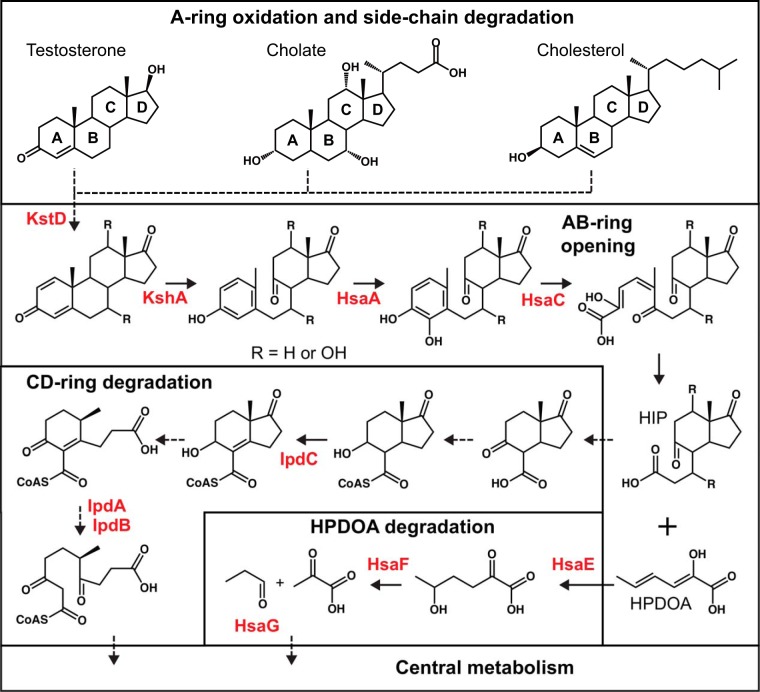
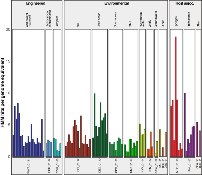
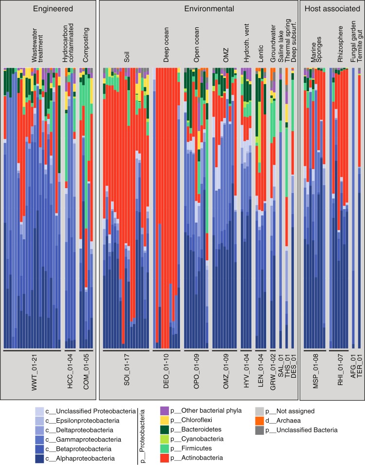
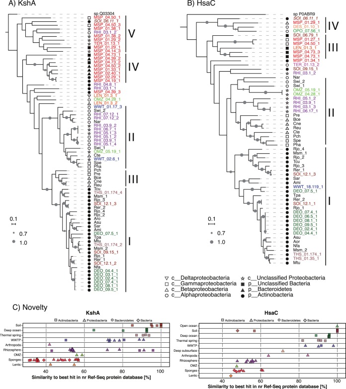
Steroids are abundant growth substrates for bacteria in natural, engineered, and host-associated environments. This study analyzed the distribution of the aerobic 9,10-seco steroid degradation pathway in 346 publically available metagenomes from diverse environments. Our results show that steroid-degrading bacteria are globally distributed and prevalent in particular environments, such as wastewater treatment plants, soil, plant rhizospheres, and the marine environment, including marine sponges. Genomic signature-based sequence binning recovered 45 metagenome-assembled genomes containing a majority of 9,10-seco pathway genes. Only Actinobacteria and Proteobacteria were identified as steroid degraders, but we identified several alpha- and gammaproteobacterial lineages not previously known to degrade steroids. Actino- and proteobacterial steroid degraders coexisted in wastewater, while soil and rhizosphere samples contained mostly actinobacterial ones. Actinobacterial steroid degraders were found in deep ocean samples, while mostly alpha- and gammaproteobacterial ones were found in other marine samples, including sponges. Isolation of steroid-degrading bacteria from sponges confirmed their presence. Phylogenetic analysis of key steroid degradation proteins suggested their biochemical novelty in genomes from sponges and other environments. This study shows that the ecological significance as well as taxonomic and biochemical diversity of bacterial steroid degradation has so far been largely underestimated, especially in the marine environment.IMPORTANCE Microbial steroid degradation is a critical process for biomass decomposition in natural environments, for removal of important pollutants during wastewater treatment, and for pathogenesis of bacteria associated with tuberculosis and other bacteria. To date, microbial steroid degradation was mainly studied in a few model organisms, while the ecological significance of steroid degradation remained largely unexplored. This study provides the first analysis of aerobic steroid degradation in diverse natural, engineered, and host-associated environments via bioinformatic analysis of an extensive metagenome data set. We found that steroid-degrading bacteria are globally distributed and prevalent in wastewater treatment plants, soil, plant rhizospheres, and the marine environment, especially in marine sponges. We show that the ecological significance as well as the taxonomic and biochemical diversity of bacterial steroid degradation has been largely underestimated. This study greatly expands our ecological and evolutionary understanding of microbial steroid degradation.

Keywords: Comamonas; Mycobacterium; Pseudomonas; Rhodococcus; cholesterol; metagenomics; sponges; steroid degradation.


Figures

FIG 1
FIG 1
Aerobic 9,10-seco degradation pathways for cholesterol, cholate, and testosterone. The steroid nucleus is degraded by oxygen-dependent opening and subsequent hydrolytic cleavage of rings A and B, leading to the formation and further degradation of (3′-propanoate)-7aβ-methylhexahydro-1,5-indanediones (HIP) and 2-hydroxyhexa-2,4-dienonic acid (HPDOA). Names of characterized steroid degradation protein families with available hidden Markov models are highlighted in red. Dashed arrows indicate multiple enzymatic reactions.
FIG 2
FIG 2
Normalized HMM hit counts of 105 metagenomes with HMM hits for all 10 steroid degradation protein families. Metagenomes are labeled using a three-letter code representing the global environment and a unique metagenome number (see Table S1A for details). Bars are color coded by global environment.
FIG 3
FIG 3
Taxonomic classification of predicted steroid degradation proteins. Shown are class-, phylum-, or domain-level assignments of predicted steroid degradation proteins in 105 analyzed metagenomes. “Other bacterial phyla” includes all phyla assigned to less than 1% of the proteins. Metagenomes are labeled using a three-letter code representing the global environment and a unique metagenome number (see Table S1A for details).
FIG 4
FIG 4
Phylogeny of (A) KshA and (B) HsaC proteins from predicted steroid degrader metagenome-assembled genomes (MAGs). Shapes represent the lowest common ancestor phylum or class classification of KshA and HsaC from MAGs and of strain taxonomy for KshA and HsaC from complete genomes. Gray circles represent bootstrap support values; only bootstrap values over 70% are shown. The scales correspond to 0.1 substitution per amino acid. KshA and HsaC proteins from MAGs that do not encode orthologs of known steroid degradation proteins are italic. The three-letter codes for steroid-degrading bacteria are as follows: Ami, Actinoplanes missouriensis 431; Aro, Amycolatopsis orientalis HCCB10007; Asu, Amycolicicoccus subflavus DQS3-9A1; Bce, Burkholderia cepacia GG4; Cte, Comamonas testosteroni CNB-2; Cne, Cupriavidus necator N-1; Msm, Mycobacterium smegmatis MC2155; Mtu, Mycobacterium tuberculosis H37Rv; Nfa, Nocardia farcinica IFM 10152; Nar, Novosphingobium aromaticivorans DSM12444; Pha, Pseudoalteromonas haloplanktis TAC125; Pre, Pseudomonas resinovorans NBRC106553; Pch, Pseudomonas sp. strain Chol1; Reu, Ralstonia eutropha H16; Rer, Rhodococcus erythropolis PR4; Rjo, Rhodococcus jostii RHA1; Sar, Salinispora arenicola CNS-205; Spe, Shewanella pealeana ATCC 700345; Swi, Sphingomonas wittichii RW1; Tcu, Thermomonospora curvata DSM43183; Tpa, Tsukamurella paurometabola DSM20162. Toluene-4-monooxygenase (GenBank accession no. Q03304.1) from Pseudomonas medocina and carboxyethylcatechol 2,3-dioxygenase (GenBank accession no. P0ABR9.1) from Escherichia coli K-12 were used as outgroups for KshA and HsaC, respectively. (C) Novelty of KshA and HsaC proteins in predicted steroid degrader MAGs. Similarity values of protein sequences to their best hit in the non-redundant RefSeq protein database are shown. Shapes represent lowest common ancestor phylum or domain classification. Protein identifications (IDs) in panels A and B and individual proteins in panel C are color coded by global environment as in Fig. 2.

References

    1. Desmond E, Gribaldo S. 2009. Phylogenomics of sterol synthesis: insights into the origin, evolution, and diversity of a key eukaryotic feature. Genome Biol Evol 1:364–381. doi: 10.1093/gbe/evp036. - DOI - PMC - PubMed
    1. Gold DA, Caron A, Fournier GP, Summons RE. 2017. Paleoproterozoic sterol biosynthesis and the rise of oxygen. Nature 543:420–423. doi: 10.1038/nature21412. - DOI - PubMed
    1. Gold DA, Grabenstatter J, de Mendoza A, Riesgo A, Ruiz-Trillo I, Summons RE. 2016. Sterol and genomic analyses validate the sponge biomarker hypothesis. Proc Natl Acad Sci U S A 113:2684–2689. doi: 10.1073/pnas.1512614113. - DOI - PMC - PubMed
    1. Zhou H, Hylemon PB. 2014. Bile acids are nutrient signaling hormones. Steroids 86:62–68. doi: 10.1016/j.steroids.2014.04.016. - DOI - PMC - PubMed
    1. Buchinger TJ, Li W, Johnson NS. 2014. Bile salts as semiochemicals in fish. Chem Senses 39:647–654. doi: 10.1093/chemse/bju039. - DOI - PubMed

Publication types

LinkOut - more resources