. 2018 Jan 31;8(1):33.
doi: 10.1038/s41398-017-0074-6.

Extended amygdala connectivity changes during sustained shock anticipation

Affiliations

Extended amygdala connectivity changes during sustained shock anticipation

Salvatore Torrisi et al. Transl Psychiatry. .

Abstract

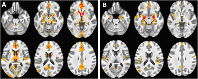
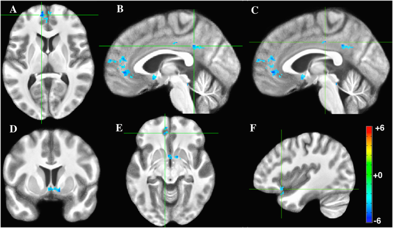
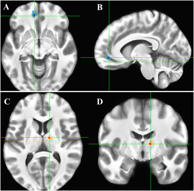
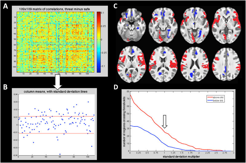
The bed nucleus of the stria terminalis (BNST) and central amygdala (CeA) of the extended amygdala are small, anatomically interconnected brain regions. They are thought to mediate responses to sustained, unpredictable threat stimuli and phasic, predictable threat stimuli, respectively. They perform these operations largely through their interconnected networks. In two previous studies, we mapped and contrasted the resting functional connectivity networks of the BNST and CeA at 7 Tesla with high resolution. This follow-up study investigates the changes in functional connectivity of these structures during sustained anticipation of electric shock. Results show that the BNST and CeA become less strongly coupled with the ventromedial prefrontal cortex (vmPFC), cingulate, and nucleus accumbens in shock threat relative to a safety condition. In addition, the CeA becomes more strongly coupled with the thalamus under threat. An exploratory, whole-brain connectivity analysis reveals that, although the BNST/CeA exhibits generally decreased connectivity, many other cortical regions demonstrate greater coupling under threat than safety. Understanding the differential network structures of these two regions and how they contribute to processing under threat will help elucidate the building blocks of the anxious state.

Trial registration: ClinicalTrials.gov NCT00047853 NCT00001360.


Conflict of interest statement

The authors declare that they have no conflict of interest.

Figures

Fig. 1
Fig. 1. Resting connectivity during safety blocks.
a BNST functional connectivity. b CeA functional connectivity. Results thresholded at p < 1 × 10−7, k = 100. Results in these and subsequent figures are overlaid on the average of subjects’ structural scans
Fig. 2
Fig. 2. BNST connectivity, threat vs. safety contrast.
a Axial slice of medial prefrontal cortex (mPFC). b Sagittal slice of posterior cingulate. c Sagittal slice of posterior cingulate. d Coronal slice of nucleus accumbens. e Axial slice of ventromedial PFC (vmPFC). f Sagittal slice of ventrolateral prefrontal cortex (vlPFC). Figure thresholded at p < 0.005, k = 54. Note that here negative t-statistics represent greater positive connectivity during the safety condition, and not negative connectivity
Fig. 3
Fig. 3. CeA connectivity, threat vs. safety contrast.
Axial slice results: a ventromedial prefrontal cortex (vmPFC). b Right ventral–anterior nucleus of the thalamus. Color scheme is the same as Fig. 2
Fig. 4
Fig. 4. Qualitative exploratory analysis.
a 109 region correlation matrix, as contrasted Threat > Safety. b Mean correlations (i.e., 4 A matrix columns collapsed) with SD lines in red. See Table 2 for regions above and below 1 SD from mean. c Ventral to dorsal axial slices of > ± 1 SD regions visualized on the brain: positive correlation regions red, negative correlation regions blue. d Thresholding in (b) and (c) was arbitrarily set at 1 SD; however, the observation that more regions were on average more positively connected during threat than safety is invariant to thresholding (red greater than blue line across thresholds: down arrow points to 1 SD point)

Similar articles

Cited by

References

    1. Lissek S, et al. Classical fear conditioning in the anxiety disorders: a meta-analysis. Behav. Res. Ther. 2005;43:1391–1424. doi: 10.1016/j.brat.2004.10.007. - DOI - PubMed
    1. Ressler KJ. Amygdala activity, fear, and anxiety: modulation by stress. Biol. Psychiatry. 2010;67:1117–1119. doi: 10.1016/j.biopsych.2010.04.027. - DOI - PMC - PubMed
    1. LeDoux Joseph E., Pine Daniel S. Using Neuroscience to Help Understand Fear and Anxiety: A Two-System Framework. American Journal of Psychiatry. 2016;173(11):1083–1093. doi: 10.1176/appi.ajp.2016.16030353. - DOI - PubMed
    1. Davis M, Walker DL, Miles L, Grillon C. Phasic vs sustained fear in rats and humans: role of the extended amygdala in fear vs anxiety. Neuropsychopharmacology. 2010;35:105–135. doi: 10.1038/npp.2009.109. - DOI - PMC - PubMed
    1. Davis M, Whalen PJ. The amygdala: vigilance and emotion. Mol. Psychiatry. 2001;6:13–34. doi: 10.1038/sj.mp.4000812. - DOI - PubMed

Publication types

MeSH terms

Associated data