

Tbx5a lineage tracing shows cardiomyocyte plasticity during zebrafish heart regeneration
- PMID: 29382818
- PMCID: PMC5789846
- DOI: 10.1038/s41467-017-02650-6
Tbx5a lineage tracing shows cardiomyocyte plasticity during zebrafish heart regeneration
Abstract
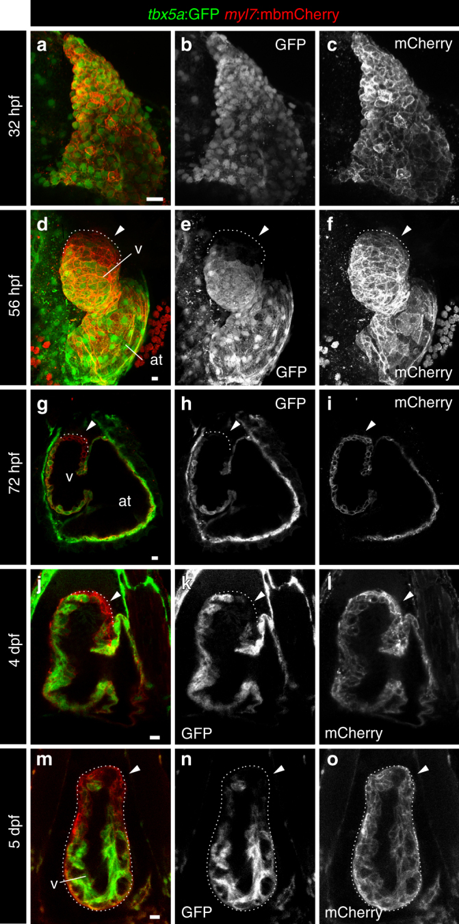
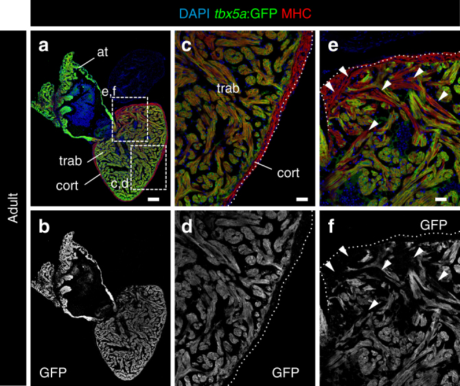
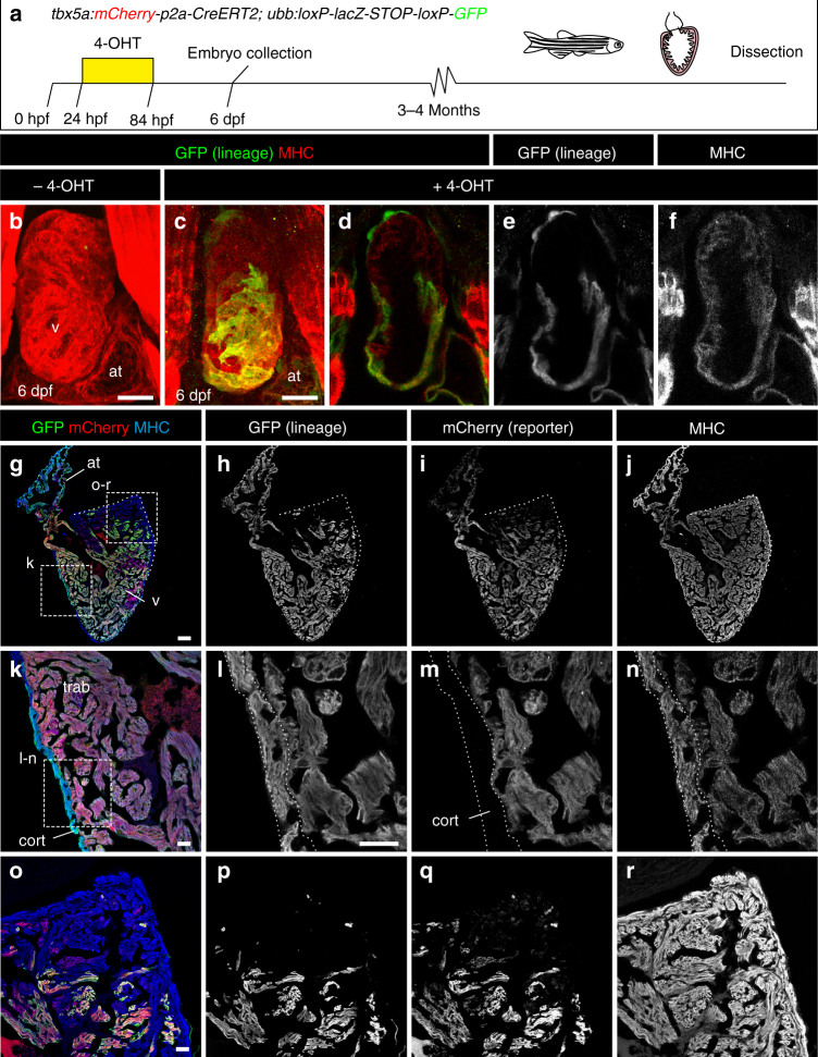
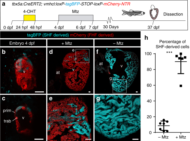
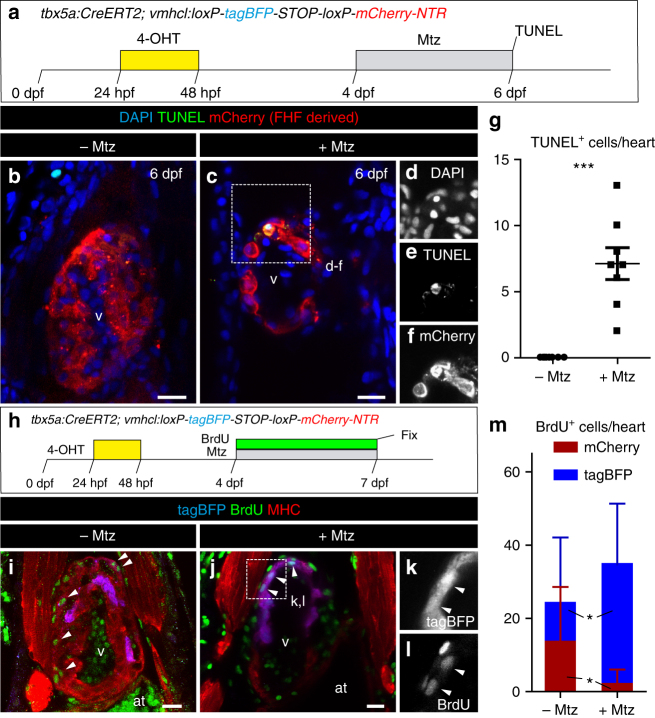
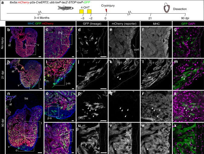
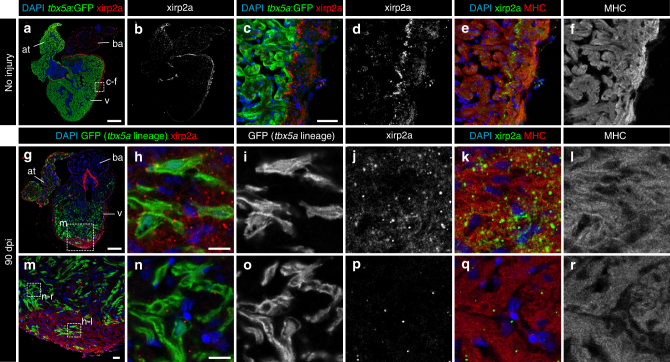
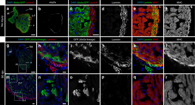
During development, mesodermal progenitors from the first heart field (FHF) form a primitive cardiac tube, to which progenitors from the second heart field (SHF) are added. The contribution of FHF and SHF progenitors to the adult zebrafish heart has not been studied to date. Here we find, using genetic tbx5a lineage tracing tools, that the ventricular myocardium in the adult zebrafish is mainly derived from tbx5a+ cells, with a small contribution from tbx5a- SHF progenitors. Notably, ablation of ventricular tbx5a+-derived cardiomyocytes in the embryo is compensated by expansion of SHF-derived cells. In the adult, tbx5a expression is restricted to the trabeculae and excluded from the outer cortical layer. tbx5a-lineage tracing revealed that trabecular cardiomyocytes can switch their fate and differentiate into cortical myocardium during adult heart regeneration. We conclude that a high degree of cardiomyocyte cell fate plasticity contributes to efficient regeneration.
Conflict of interest statement
The authors declare no competing financial interests.
Figures


References
Publication types
MeSH terms
Substances
Grants and funding
LinkOut - more resources
Full Text Sources
Other Literature Sources
Molecular Biology Databases
Research Materials
