. 2018 Jan 30;9(1):430.
doi: 10.1038/s41467-018-02897-7.

Gimap5-dependent inactivation of GSK3β is required for CD4+ T cell homeostasis and prevention of immune pathology

Affiliations

Gimap5-dependent inactivation of GSK3β is required for CD4+ T cell homeostasis and prevention of immune pathology

Andrew R Patterson et al. Nat Commun. .

Abstract

GTPase of immunity-associated protein 5 (Gimap5) is linked with lymphocyte survival, autoimmunity, and colitis, but its mechanisms of action are unclear. Here, we show that Gimap5 is essential for the inactivation of glycogen synthase kinase-3β (GSK3β) following T cell activation. In the absence of Gimap5, constitutive GSK3β activity constrains c-Myc induction and NFATc1 nuclear import, thereby limiting productive CD4+ T cell proliferation. Additionally, Gimap5 facilitates Ser389 phosphorylation and nuclear translocation of GSK3β, thereby limiting DNA damage in CD4+ T cells. Importantly, pharmacological inhibition and genetic targeting of GSK3β can override Gimap5 deficiency in CD4+ T cells and ameliorates immunopathology in mice. Finally, we show that a human patient with a GIMAP5 loss-of-function mutation has lymphopenia and impaired T cell proliferation in vitro that can be rescued with GSK3 inhibitors. Given that the expression of Gimap5 is lymphocyte-restricted, we propose that its control of GSK3β is an important checkpoint in lymphocyte proliferation.


Conflict of interest statement

The authors declare no competing financial interests.

Figures

Fig. 1
Fig. 1
Loss of Gimap5 impairs CD4+ T cell survival and iTreg cell induction. Ten-week-old control Gimap5WT/WT;Rag2/;OT-II and Gimap5sph/sph;Rag2/;OT-II mice received either normal water or water containing 1 mg/mL ovalbumin ad libitum during a 5-week period. At 15 weeks of age, a the number of splenic CD4+ T cells and b the percentage of naïve (CD44lo;CD62hi) and memory-like (CD44hi;CD62lo) CD4+ T cells was determined using flow cytometry. c Frequency of iTregs (CD4+CD25+) in the mesenteric lymph nodes (MLN) of vehicle- and OVA-treated mice (n = 4). d Survival of peripheral (splenic) CD4+ T cells ex vivo in the presence of IL-7 (5 ng/mL) as determined by live/dead staining and flow cytometry (n = 3). Data represent mean values + SD of samples from individual mice; statistical significance is determined by Student’s two-tailed test
Fig. 2
Fig. 2
Impaired CD4+ T cell proliferation is associated with increased GSK3 activity. a Immunoblot analysis of c-Myc expression in total lysates of CD4+ T cells from WT and Gimap5sph/sph mice stimulated with αCD3/αCD28 or during resting conditions. b Myc mRNA levels in resting and αCD3/αCD28-activated (24 h) CD4+ T cells. Data represent mean expression ± SD relative to unstimulated WT cells (n = 6). c Phosphorylation c-Myc (T58) after 24 h of αCD3/αCD28 stimulation. Proteasomal inhibitor MG132 was added after 20 h of stimulation. Ratio of p-c-Myc (T58) to total c-Myc in MG132-treated Gimap5sph/sph CD4+ T cells relative to WT (n = 5). d C-Myc expression in WT and Gimap5sph/sph CD4+ T cells stimulated for 24 h with αCD3/αCD28 ± 2.5 mM LiCl. e Proliferation of WT and Gimap5sph/sph CD4+ T cells in the presence/absence of αCD3/αCD28 and/or GSK3 inhibitors BIO (100 nM) or LiCl (2.5 mM) as measured by CFSE dilution after 3 days. Experiments were repeated five times and representative plots from a single experiment are shown. f Representative images of NFATc1 localization in resting and αCD3/αCD28-activated WT and Gimap5sph/sph CD4+ T cells. Bright detail similarity quantification of NFATc1 nuclear localization upon stimulation (g) and in the presence/absence of BIO (h) for 4 h (n = 6). Graphs depict mean values ± SEM. ImageStream data represent average values of >500 CD4+ T cells per sample; all experiments were performed at least three times. Statistical significance is determined by Student’s two-tailed test. BF bright field
Fig. 3
Fig. 3
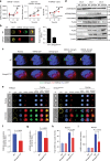
Loss of Gimap5 results in impaired inactivation of GSK3β. a Association of GSK3β with vesicles in WT and Gimap5sph/sph CD4+ T cells as exemplified by GSK3β intensity, vesicle number, and vesicle size of GSK3β spots 6 and 24 h after αCD3/αCD28 activation. b Bright detail similarity analysis (ImageStream) of activated CD4+T cells from WT and Gimap5sph/sph mice show colocalization between Gimap5 and GSK3β. c Representative Z-stacks of WT and Gimap5sph/sph CD4+ T cells stimulated for 24 h with αCD3/αCD28. d Immunoblot analysis of WT and Gimap5sph/sph CD4+ T cells stimulated with αCD3/αCD28 depicting total and phosphorylated protein levels of GSK3β (P-Ser9 and P-Ser389), p38, and p53. e–i Localization of total and phospho-GSK3β (Ser389) in WT or Gimap5sph/sph CD4+ T cells after 2 days stimulation with αCD3/αCD28 (n = 3). f Nuclear localization and h expression of total GSK3β. g Nuclear localization and i expression of p-GSK3β (Ser389). Hatched bars represent resting and solid bars represent CD3/CD28-activated CD4+ T cells. Graphs depict mean values ± SD. ImageStream data represent average values of >500 CD4+ T cells per sample. All experiments were performed at least three times. Statistical significance is determined by Student’s two-tailed test. BF bright field
Fig. 4
Fig. 4
Loss of Gimap5 causes increased DNA damage in activated CD4+ T cells. a γH2AX expression in live WT or Gimap5sph/sph CD4+ T cells following αCD3/αCD28 stimulation. b Survival as determined by viability stain of WT or Gimap5sph/sph CD4+ T cells after αCD3/αCD28 stimulation (n = 4). Effect of lithium on γH2AX expression in WT or Gimap5sph/sph CD4+ T cells, after 3 days stimulation with αCD3/αCD28. Plots depict live cells (c, d), while bar graphs (d) represent mean values ± SD (n = 6). All experiments were performed at least three times. Statistical significance is determined by Student’s two-tailed test
Fig. 5
Fig. 5
GSK3 inhibition improves lymphocyte survival and prevents immunopathology. LiCl treatment of Gimap5sph/sph mice in vivo starting at 3 weeks of age and analyzed at 7–8 weeks of age, rescues CD4+ T cell (a) and B cell (b) survival. c Reduced frequency of CD4+ T cells undergoing lymphopenia-induced proliferation (CD44hi;CD62lo) upon LiCl treatment of Gimap5sph/sph mice in vivo. d Suppressive capacity of regulatory T cells (CD4+CD25hi) isolated from vehicle or LiCl-treated WT and Gimap5sph/sph mice. Data represents mean ± SD and is representative to two independent experiments (n = 3). Reduced colitis as defined by disease score (e) and based on colon histology (f) in 7–8-week-old Gimap5sph/sph mice following LiCl treatment (3.5 weeks) in vivo. g Reduced liver pathology in Gimap5sph/sph treated with LiCl. Data represent mean values ± SD from 6 mice per group at 7–8 weeks of age; histology is a representative depiction of disease severity. Scale bar is 50 μm. Statistical significance is determined by ANOVA followed by Sidak’s multiple comparisons test
Fig. 6
Fig. 6
GSK3β deletion in CD4+ T cells improves CD4+ T cell survival and prevents colitis. a Tamoxifen treatment of Gimap5sph/sph; Gsk3bfl/fl; Cd4cre-ert2 mice starting at 3 weeks of age selectively rescues splenic CD4+ T cell survival, b while maintaining overall CD4+ T cell quiescence. Reduced colitis in Gimap5sph/sph; Gsk3bfl/fl; Cd4cre-ert2 mice treated with tamoxifen as determined by disease scores (c) based on histology (d). Data represent mean values ± SEM from at least 6 mice per group at 8 weeks of age; histology is a representative depiction of disease severity. Scale bar is 50 μm. Statistical significance is determined by ANOVA followed by Sidak’s multiple comparisons test
Fig. 7
Fig. 7
A human loss-of-function mutation in GIMAP5 results in a similar T cell deficiency. a Whole-exome sequencing uncovered a homozygous variant (SNP: rs72650695), causing an L204P amino acid change in GIMAP5, resulting in complete loss of GIMAP5 protein expression in T cells. b Impaired expansion of CD3+ T cells from GIMAP5/ patient compared to heterozygous control cells following stimulation with PHA (4 days) and IL-2 (days 4–10). The proliferation capacity is restored in the presence of LiCl. Experiment is representative of three independent experiments from samples obtained several months apart. Immunoblot analysis of control and GIMAP5/ CD3+ T cells shows reduced c-Myc expression (c) at resting conditions and (d) after 2-day restimulation with αCD3/αCD28 ± 5 mM LiCl. Immunoblot was repeated twice with an identical outcome
Fig. 8
Fig. 8
Human GIMAP5/ T cells show impaired GSK3β sequestration and increased DNA damage. ad Colocalization and spot analysis of GIMAP5+, GSK3β+, and CD107+ vesicles in primary CD4+ T cells isolated from healthy controls at a resting state (day 0) or after αCD3/αCD28 stimulation for 1–2 days (representative images from day 1 shown) using ImageStream analysis. eh ImageStream analysis of GSK3β-specific vesicle association in control or GIMAP5/ patient CD4+ T cells restimulated with αCD3/αCD28 after primary expansion. Data depicts e GSK3β+ spot number, f GSK3β intensity therein, and g spot area in live CD4+ T cells at 0, 6, or 24 h of αCD3/αCD28-restimulation. Data represent mean values ± SD. h Representative images of GSK3β vesicular association in control and GIMAP5/ CD4+ T cells taken using a 60× objective. Analysis of DNA damage response (γH2AX) in control of GIMAP5sph/sph CD4+ T cells after i 1–3 days or j 2 days restimulation with αCD3/αCD28. Data represents mean values of a single experiment performed in duplicate and repeated twice. ImageStream data represent average values of >500 CD4+ T cells per experiment. BF bright field
Fig. 9
Fig. 9
Gimap5 is a critical regulator of GSK3β during T cell activation. Gimap5 controls regulation of GSK3β in T cells through vesicular sequestration and affects both the (1) (early) transcriptional program required for T cell growth, and (2) the late stage nuclear accumulation of P-Ser389 GSK3β required for the DNA damage response during cycling. Gimap5-deficient CD4+ T cells, fail to inhibit GSK3β leading to a failed transcriptional program and increased DNA damage during T cells proliferation

Similar articles

Cited by

  • GIMAP6 is required for T cell maintenance and efficient autophagy in mice.
    Pascall JC, Webb LMC, Eskelinen EL, Innocentin S, Attaf-Bouabdallah N, Butcher GW. Pascall JC, et al. PLoS One. 2018 May 2;13(5):e0196504. doi: 10.1371/journal.pone.0196504. eCollection 2018. PLoS One. 2018. PMID: 29718959 Free PMC article.
  • When ceramides meet immune senescence, a GIMAP5 connection.
    Gope A, Mauro C. Gope A, et al. Nat Immunol. 2024 Feb;25(2):196-197. doi: 10.1038/s41590-023-01736-2. Nat Immunol. 2024. PMID: 38263464 No abstract available.
  • GIMAP5 deficiency reveals a mammalian ceramide-driven longevity assurance pathway.
    Park AY, Leney-Greene M, Lynberg M, Gabrielski JQ, Xu X, Schwarz B, Zheng L, Balasubramaniyam A, Ham H, Chao B, Zhang Y, Matthews HF, Cui J, Yao Y, Kubo S, Chanchu JM, Morawski AR, Cook SA, Jiang P, Ravell JC, Cheng YH, George A, Faruqi A, Pagalilauan AM, Bergerson JRE, Ganesan S, Chauvin SD, Aluri J, Edwards-Hicks J, Bohrnsen E, Tippett C, Omar H, Xu L, Butcher GW, Pascall J, Karakoc-Aydiner E, Kiykim A, Maecker H, Tezcan İ, Esenboga S, Heredia RJ, Akata D, Tekin S, Kara A, Kuloglu Z, Unal E, Kendirli T, Dogu F, Karabiber E, Atkinson TP, Cochet C, Filhol O, Bosio CM, Davis MM, Lifton RP, Pearce EL, Daumke O, Aytekin C, Şahin GE, Aksu AÜ, Uzel G, Koneti Rao V, Sari S, Dalgıç B, Boztug K, Cagdas D, Haskologlu S, Ikinciogullari A, Schwefel D, Vilarinho S, Baris S, Ozen A, Su HC, Lenardo MJ. Park AY, et al. Nat Immunol. 2024 Feb;25(2):282-293. doi: 10.1038/s41590-023-01691-y. Epub 2024 Jan 3. Nat Immunol. 2024. PMID: 38172257 Free PMC article.
  • The Role of GSK3β in T Lymphocytes in the Tumor Microenvironment.
    Dimou A, Syrigos KN. Dimou A, et al. Front Oncol. 2020 Jul 24;10:1221. doi: 10.3389/fonc.2020.01221. eCollection 2020. Front Oncol. 2020. PMID: 32850361 Free PMC article. Review.
  • Identification and validation of GIMAP family genes as immune-related prognostic biomarkers in lung adenocarcinoma.
    Zhang Y, Liu S, Liu D, Zhao Z, Song H, Peng K. Zhang Y, et al. Heliyon. 2024 Jun 15;10(12):e33111. doi: 10.1016/j.heliyon.2024.e33111. eCollection 2024 Jun 30. Heliyon. 2024. PMID: 38948046 Free PMC article.

References

    1. Hellquist A, et al. The human GIMAP5 gene has a common polyadenylation polymorphism increasing risk to systemic lupus erythematosus. J. Med. Genet. 2007;44:314–321. doi: 10.1136/jmg.2006.046185. - DOI - PMC - PubMed
    1. Lim MK, et al. IAN5 polymorphisms are associated with systemic lupus erythematosus. Lupus. 2009;18:1045–1052. doi: 10.1177/0961203309106830. - DOI - PubMed
    1. Shin JH, et al. IA-2 autoantibodies in incident type I diabetes patients are associated with a polyadenylation signal polymorphism in GIMAP5. Genes Immun. 2007;8:503–512. doi: 10.1038/sj.gene.6364413. - DOI - PubMed
    1. Heinonen MT, et al. GIMAP GTPase family genes: potential modifiers in autoimmune diabetes, asthma, and allergy. J. Immunol. 2015;194:5885–5894. doi: 10.4049/jimmunol.1500016. - DOI - PMC - PubMed
    1. Aksoylar HI, Lampe K, Barnes MJ, Plas DR, Hoebe K. Loss of immunological tolerance in Gimap5-deficient mice is associated with loss of Foxo in CD4+T Cells. J. Immunol. 2012;188:146–154. doi: 10.4049/jimmunol.1101206. - DOI - PMC - PubMed

Publication types

MeSH terms