. 2018 Jan 30;8(1):1916.
doi: 10.1038/s41598-018-19530-8.

APLP1 is endoproteolytically cleaved by γ-secretase without previous ectodomain shedding

Affiliations

APLP1 is endoproteolytically cleaved by γ-secretase without previous ectodomain shedding

Linda Schauenburg et al. Sci Rep. .

Abstract

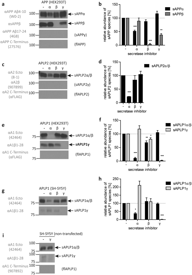
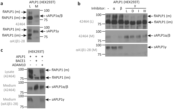
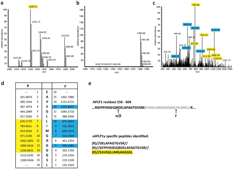
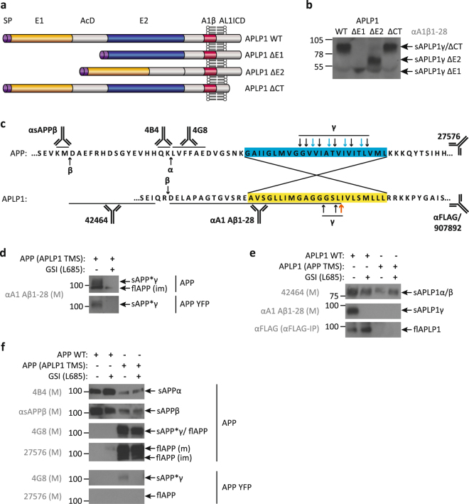
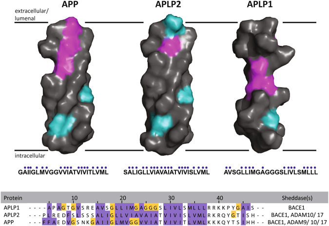
Regulated intramembrane proteolysis of the amyloid precursor protein (APP) and its homologs, the APP like proteins APLP1 and APLP2, is typically a two-step process, which is initiated by ectodomain-shedding of the substrates by α- or β-secretases. Growing evidence, however, indicates that the cleavage process for APLP1 is different than for APP. Here, we describe that full-length APLP1, but not APP or APLP2, is uniquely cleaved by γ-secretase without previous ectodomain shedding. The new fragment, termed sAPLP1γ, was exclusively associated with APLP1, not APP, APLP2. We provide an exact molecular analysis showing that sAPLP1γ was uniquely generated by γ-secretase from full-length APLP1. Mass spectrometry analysis showed that the sAPLP1γ fragment and the longest Aβ-like peptide share the C-terminus. This novel mechanism of γ-secretase action is consistent with an ϵ-cut based upon the nature of the reaction in APP. We further demonstrate that the APLP1 transmembrane sequence is the critical determinant for γ-shedding and release of full-length APLP1. Moreover, the APLP1 TMS is sufficient to convert larger type-I membrane proteins like APP into direct γ-secretase substrates. Taken together, the direct cleavage of APLP1 is a novel feature of the γ-secretase prompting a re-thinking of γ-secretase activity modulation as a therapeutic strategy for Alzheimer disease.


Conflict of interest statement

The authors declare that they have no competing interests.

Figures

Figure 1
Figure 1
Western blot analysis of APP, APLP2 and APLP1 processing in HEK293T and SH-SY5Y cells and quantitative analyses of detected fragments. Western blot analysis of soluble fragments in the media of HEK293T cells overexpressing APP (a), APLP2-FLAG (c) and APLP1-FLAG (e) as well as APLP1-overexpressing SH-SY5Y cells (g) and of non-transfected SH-SY5Y cells with or without γ-secretase inhibition after immunoprecipitation from conditioned medium with αAPLP1ecto antibody and detection with the antibodies 42464, αA1β1–28 and 907892 (i). Conditioned media were analyzed using specific antibodies for the soluble ectodomains (APLP1–42464; APLP2–8–1; APP – W0–2 or αsAPPβ), the Aβ-region (APLP1 – αA1β1–28; APLP2–907899; APP – 4G8) or the C-terminus (APLP1/APLP2 – αFLAG; APP – 27576). Transfected cells were treated with α- (GM6001, Calbiochem), β- (β-secretase inhibitor IV, Calbiochem) or γ-secretase inhibitor (L-685,458 Calbiochem). All respective bands were quantified, normalized against the untreated control (100%) and statistically analyzed by one-sample t-test vs. 100% with Bonferroni correction (*p < 0.05, **p < 0.01, ***p < 0.001, (b,d,f,h)). Displayed are representative Western blots of at least three independent experiments. The actual number of repeats and representative full-length blots are given and presented in Supplementary Figure 1, respectively.
Figure 2
Figure 2
Western blot analysis of soluble and full-length APLP1 species. Conditioned medium (M) or lysate (L) of HEK293T cells overexpressing APLP1 was analyzed with the anti APLP1 antibodies 42464 and αA1β1–28 to display differences in size between sAPLP1γ, fl-APLP1 and sAPLP1α/β and between mature (m) and immature (im) full-length APLP1 (a). In HEK293T cells overexpressing APLP1 treated with α-, β- or different γ-secretase inhibitors (α – GM6001; β – β-secretase-inhibitor IV; γ/ L – L-685,458; D – DAPT; I – GSI I; III – GSI III), full-length and secreted forms were analyzed in L and M with the antibodies as indicated (b). HEK293T cells were co-transfected with APLP1 and BACE1 or ADAM10 and full-length and secreted forms analyzed in L and M with the indicated antibodies (c). Displayed are representative Western blots of two independent experiments. Full-length blots of both experiments are presented in Supplementary Figures 2 and 3.
Figure 3
Figure 3
LC-MS analysis to identify the sAPLP1γ C-terminus. Tryptic peptide spectrum of sAPLP1γ (a) and sAPLP1α/β (b) at about 1400 Da. The peak at 1430.75 only appears in sAPLP1γ samples. MS/MS analysis of the sAPLP1γ peptide at m/z = 1430.75 Da (c) and comparison with expected b and y sequence fragments of the APLP1 peptide EAVSGLLIMGAGGGSL (d). The peptides highlighted in yellow (b) and blue (y) were detected. APLP1 sequence 556–604 with indicated α-, β- and γ-cleavage sites (TMS in grey) and list of tryptic and semi-tryptic peptides from that sequence found in sAPLP1γ samples (e).
Figure 4
Figure 4
Soluble processing products of APLP1 deletion mutants and APLP1-APP chimeras. Schematic representation of the APLP1 domains and APLP1 deletion mutants analyzed: SP – signal peptide; E1 and E2 – two conserved regions of the ectodomain; AcD – acidic stretch linker region between E1 and E2; A1β – amyloid beta-like sequence of APLP1; AL1ICD – APLP1 intracellular domain; CT – C-terminus (a). Conditioned media of HEK293T cells overexpressing the APLP1 deletion mutants, detection of sAPLP1γ species with the polyclonal antibody αA1β1–28 (b). Amino-acid sequences of APP and APLP1 around the transmembrane sequence (TMS – yellow or blue box), indicated are the cleavage sites of α-, β- and γ-secretase and the recognition sites of the antibodies used (c). For the creation of the chimeric proteins the TMS of APLP1 was inserted into the APP sequence and vice versa to replace the respective TMS. Analysis of the conditioned media of HEK293T cells overexpressing WT or chimeric APP and APLP1. Soluble species and full-length forms in the conditioned media of HEK 293T cells overexpressing APP WT and APP (APLP1 TMS) with or without C-terminal YFP tag were detected in the presence or absence of the γ-secretase inhibitor L – L-685,458 (L685) with the APLP1-specific antibody αA1β1–28 (d), APLP1 WT and APLP1 (APP TMS) with the antibodies 42464 and αA1β1–28 as well as αFLAG antibody after IP against the C-terminal FLAG tag (e) and with the APP-specific antibodies 4B4, αsAPPβ, 4G8 and 27576 (f). Displayed are representative Western blots of at least two independent experiments. Full-length blots are presented in Supplementary Figures 4 and 5.
Figure 5
Figure 5
Molecular model of the APLP1 TMS within the plasma membrane. The APLP1 TMS has a unique sequence and structural flexibility within the membrane: side-view of 3D surface representations of the putative transmembrane sequences of APP family members, assuming ideal α-helices. Hydrophobic amino acids are colored in gray, polar uncharged amino acids in cyan, and glycine in magenta. The area around the respective TMS of APP and APLPs (hydrophobic amino acids are highlighted in purple, glycines in yellow) sequences were aligned using Clustal Ω and JalView. Sheddases which are known to cleave the respective substrate are listed (ADAMs; BACE1).

References

    1. Abolmaali, B., Taylor, H. V. & Weser, U. Evolutionary aspects of copper binding centers in copper proteins, in Structure & Bonding; Bioinorganic chemistry; Trace element evolution from anaerobes to aerobes, Vol. 91. (ed. R.J.P. Williams) 91–190 (John Wiley, Berlin Heidelberg; 1998).
    1. Coulson EJ, Paliga K, Beyreuther K, Masters CL. What the evolution of the amyloid protein precursor supergene family tells us about its function. Neurochem Int. 2000;36:175–184. doi: 10.1016/S0197-0186(99)00125-4. - DOI - PubMed
    1. Wasco W, et al. Identification of a mouse brain cDNA that encodes a protein related to the Alzheimer disease-associated amyloid beta protein precursor. Proceedings of the National Academy of Sciences of the United States of America. 1992;89:10758–10762. doi: 10.1073/pnas.89.22.10758. - DOI - PMC - PubMed
    1. Wasco W, et al. Isolation and characterization of APLP2 encoding a homologue of the Alzheimer’s associated amyloid beta protein precursor. Nat Genet. 1993;5:95–100. doi: 10.1038/ng0993-95. - DOI - PubMed
    1. Heber S, et al. Mice with combined gene knock-outs reveal essential and partially redundant functions of amyloid precursor protein family members. J Neurosci. 2000;20:7951–7963. - PMC - PubMed

Publication types

MeSH terms

Substances

Grants and funding