. 2018 Mar;17(1):95-106.
doi: 10.1007/s12663-017-1019-6. Epub 2017 May 17.

Chronic Recurrent Temporomandibular Joint Dislocation: A Comparison of Various Surgical Treatment Options, and Demonstration of the Versatility and Efficacy of the Dautrey's Procedure

Affiliations

Chronic Recurrent Temporomandibular Joint Dislocation: A Comparison of Various Surgical Treatment Options, and Demonstration of the Versatility and Efficacy of the Dautrey's Procedure

Priya Jeyaraj. J Maxillofac Oral Surg. 2018 Mar.

Abstract

Introduction: There has been a changing trend of treating temporomandibular joint subluxation, which range from conservative non-surgical measures to various soft and hard tissue surgical procedures aimed at either augmenting or restricting the condylar path.

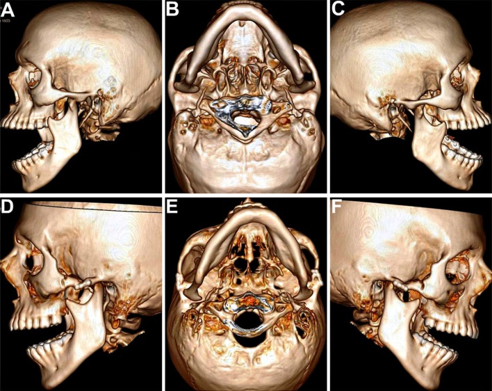
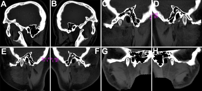
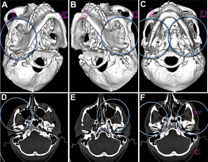
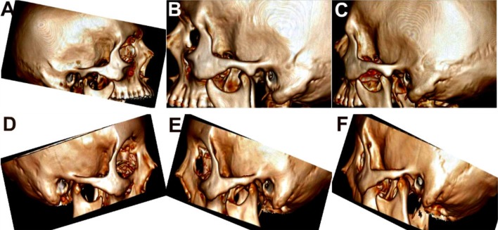
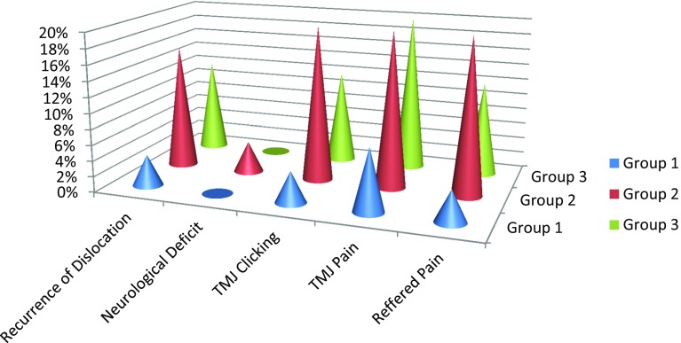
Aim: This study was aimed at comparing the efficacy of three major surgical treatment modalities: condylar obstruction creation, obstruction removal and anti-translatory procedures. Also, the location, anatomy and morphology of the TMJs pre- and post-surgery were evaluated and compared using radiographs, sagittal and 3-D Computed Tomographic scans.

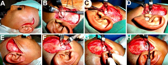
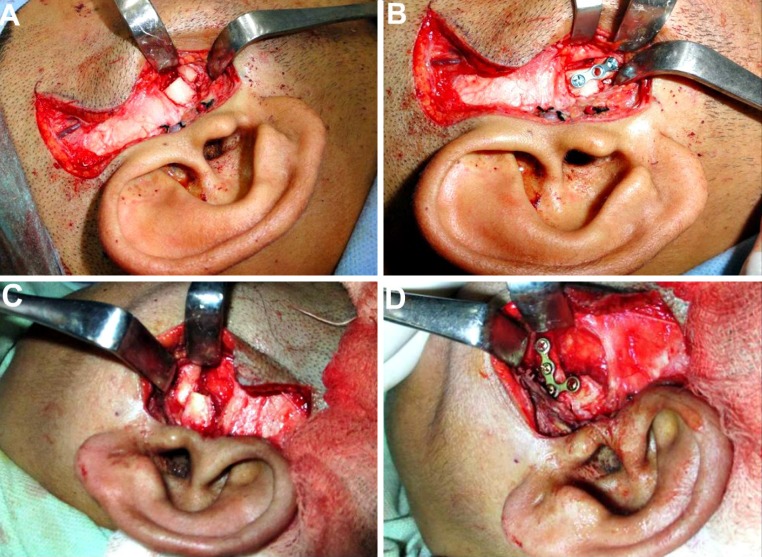
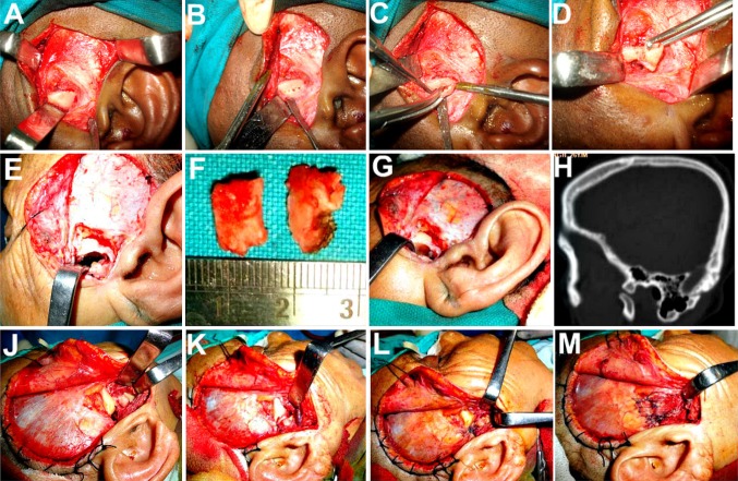
Materials and methods: A 6-year study was carried out on seventy-five patients of various age groups. Twenty-five were operated by the Dautrey's procedure, 25 by articular eminectomy alone and the remaining 25 by eminectomy followed by meniscal plication and tethering. The distribution of patients in the three groups was random. Effectiveness of the surgical procedure and incidence of complications including recurrence were carefully compiled and compared between the three groups.

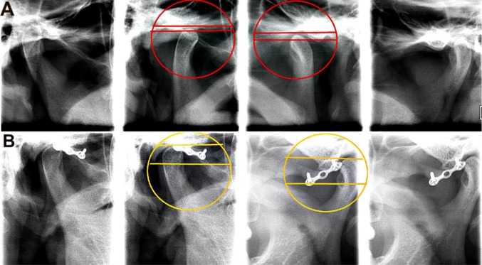
Results and conclusion: Dautrey's procedure yielded more gratifying and stable results, leading to a successful and permanent correction of chronic recurrent dislocation of the TMJs, with practically nil complications, thus demonstrating it to be an extremely safe, effective and versatile technique, making the joints function normally and securing sufficient volume of mouth opening. There was observed an average increase in articular tubercle height by 3.65 mm and a mean anterior shift of its lowest point by 4.5 mm following the Dautrey's procedure, which were statistically significant findings. The upper age limit to carry out the Dautrey's procedure can be safely taken up to 45 years.

Keywords: Arthroplasty; Capsulorrhaphy; Dautrey’s procedure; Eminectomy; Hypermobility; Meniscal plication; Subluxation.


Conflict of interest statement

Compliance with Ethical StandardsThe author of this article has not received any research grant, remuneration, or speaker honorarium from any company or committee whatsoever, and neither owns any stock in any company. The author declares that she does not have any conflict of interest.All procedures performed on the patients (human participants) involved were in accordance with the ethical standards of the institution and/or national research committee, as well as with the 1964 Helsinki declaration and its later amendments and comparable ethical standards.This article does not contain any new studies with human participants or animals performed by the author.Informed consent was obtained from all the individual participants in this study.

Figures

Fig. 1
Fig. 1
a, e Ivy and Blair’s modified preauricular incision, used to expose the TMJ and root of the zygomatic arch. b–d, f–h Osteotomy of the zygomatic arch followed by its downfracture, repositioning beneath the articular eminence and fixation using titanium minibone plate
Fig. 2
Fig. 2
a, c Care is taken to push the downfractured arch, well medially to engage as much of the mediolateral width of the condyle as possible. b, d Straight or ‘L’-shaped titanium minibone plates and screws used for fixation of the proximal arch segment in the most optimal position
Fig. 3
Fig. 3
High-resolution spiral CT scans with 3-D reconstruction. a–c Preoperative scans showing excessive anterior translation and dislocation of the condyles out of the glenoid fossae and well past the anterior slope of the articular eminences, in the open mouth position, bilaterally. d–g Postoperative scans showing restriction in forward translation of the condyles, with the condylar heads successfully restrained within the glenoid fossae by the taller neo-eminences, following their augmentation by the downfractured zygomatic arches following the Dautrey’s procedure
Fig. 4
Fig. 4
a, b Sagittal sections of CT scans showing forward dislocation of the condylar heads out of the glenoid fossae preoperatively, upon mouth opening. c–e, f–h Following the Dautrey’s procedure, the condylar heads are seen well restrained within the fossae by the augmented eminences. The downfractured arches with the fixation implants in situ are clearly seen, with the eminences being relocated more inferiorly and anteriorly. There was observed an average increase in articular tubercle height by 3.7 mm and a mean anterior shift of the lowest point of the articular tubercle by 4.5 mm following the Dautrey’s procedure
Fig. 5
Fig. 5
Simple transcranial radiographs can also be used to assess the increase in height of the neo-eminence achieved following the Dautrey’s procedure, by comparing the distance between 2 lines, the upper line drawn through the tip of the condylar head and the lower one through the tip of the articular eminence
Fig. 6
Fig. 6
1 year postoperative CT scans showing a good bony union at both, the distal fractured end of the arch as well as at the proximal augmented articular eminence region
Fig. 7
Fig. 7
a–g Bilateral Articular eminectomy, a procedure entailing removal of the convex ridge of crest affording freedom of movement to the condyle and disk, making the joint a self reducing one. h Preoperative CT scan to gauge the degree of pneumatization of the eminence so as to prevent a possible perforation into the middle cranial fossa with a resultant CSF leak or temporal lobe exposure. i–l Eminectomy combined with meniscal plication and tethering. i, j Anteriorly displaced articular disk and condylar head, repositioned back in the glenoid fossa. k, l Lateral edge of the meniscus grasped and sutured to the lax bilaminar retrodiscal tissues behind, and to the temporal fascia and muscle above, thus preventing it from slipping forward and indirectly restraining the condyle as well
Fig. 8
Fig. 8
Preoperative (a–c) and postoperative (d–f) 3-D CT scans showing a successful elimination of the convex crestal ridge from the condylar path, making the joint a self reducing one, following bilateral eminectomy
Fig. 9
Fig. 9
Postoperative mouth opening (Group 1)
Fig. 10
Fig. 10
Postoperative mouth opening (Group 2)
Fig. 11
Fig. 11
Postoperative mouth opening (Group 3)
Fig. 12
Fig. 12
Box and Whisker plot diagram showing distribution of decrease in IIO (in millimeters) among all three groups, at the end of 6 months following surgery
Fig. 13
Fig. 13
Comparison of incidence of complications post-surgery, seen in the three Groups (%age)

Similar articles

Cited by

References

    1. Helman J, Laufer D, Minkov B, Gutman D. Eminectomy as surgical treatment for chronic mandibular dislocations. Int J Oral Surg. 1984;47:179–184. - PubMed
    1. Babatunde BO. Evaluation of the mechanism and principles of management of temporomandibular joint dislocation. Systematic review of literature and a proposed new classification of temporomandibular joint dislocation. Head Face Med. 2011;7:10–14. doi: 10.1186/1746-160X-7-10. - DOI - PMC - PubMed
    1. Bhandari S, Swain M, Dewoolkar LV. Temporomandibular joint dislocation after laryngeal mask airway insertion. Internet J Anesthesiol. 2008;16:1–4.
    1. Bauss O, Sadat-Khonsari R, Fenske C, Engelke W, Schwestka-Polly R. Temporomandibular joint dysfunction in Marfan syndrome. Oral Surg Oral Med Oral Pathol Oral Radiol Endod. 2004;97:592–598. doi: 10.1016/j.tripleo.2003.10.024. - DOI - PubMed
    1. Undt G, Weichselbraun A, Wagner A, Kermer C, Rasse M. Recurrent mandibular dislocation under neuroleptic drug therapy, treated by bilateral eminectomy. J Craniomaxillofac Surg. 1996;24:184–186. doi: 10.1016/S1010-5182(96)80054-0. - DOI - PubMed

LinkOut - more resources