Review
. 2018 Jan 18:10:1758834017742575.
doi: 10.1177/1758834017742575. eCollection 2018.

Radiation effects on antitumor immune responses: current perspectives and challenges

Affiliations
Review

Radiation effects on antitumor immune responses: current perspectives and challenges

Thomas Walle et al. Ther Adv Med Oncol. .

Abstract

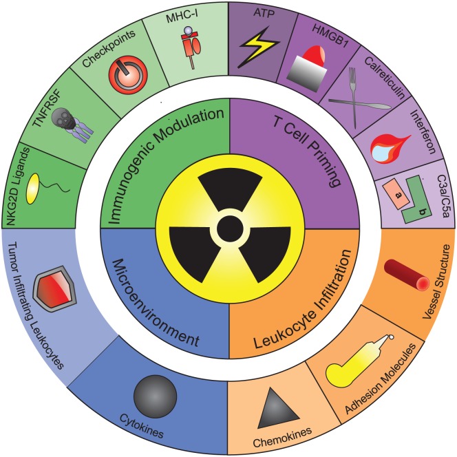
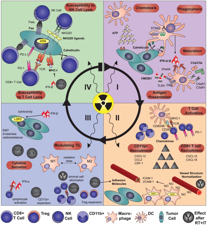
Radiotherapy (RT) is currently used in more than 50% of cancer patients during the course of their disease in the curative, adjuvant or palliative setting. RT achieves good local control of tumor growth, conferring DNA damage and impacting tumor vasculature and the immune system. Formerly regarded as a merely immunosuppressive treatment, pre- and clinical observations indicate that the therapeutic effect of RT is partially immune mediated. In some instances, RT synergizes with immunotherapy (IT), through different mechanisms promoting an effective antitumor immune response. Cell death induced by RT is thought to be immunogenic and results in modulation of lymphocyte effector function in the tumor microenvironment promoting local control. Moreover, a systemic immune response can be elicited or modulated to exert effects outside the irradiation field (so called abscopal effects). In this review, we discuss the body of evidence related to RT and its immunogenic potential for the future design of novel combination therapies.

Keywords: PD-1; abscopal; brachytherapy; checkpoint inhibitors; immunogenic cell death; immunotherapy; radiotherapy.


Conflict of interest statement

Conflict of interest statement: IM has received research support and personal fees from Bristol-Myers Squibb, Roche, AstraZeneca, Bayer, Lilly, Genmab, Incyte, Alligator, Merck-Serono, Pfizer, MSD. The rest of the authors declare no conflicts of interest.

Figures

Figure 1.
Figure 1.
Principles of the radiation-induced immune response. The effects of RT on the immune system are conceptualized in four major organizing principles (inner circle): (a) the priming of TA-specific T cells; (b) leukocyte infiltration into the tumor tissue; (c) changes in the immunosuppressive TME; and (d) immunogenic modulation of the tumor cell phenotype, leading to increased sensitivity of irradiated tumor cells to lymphocyte-mediated lysis. The mechanisms involved in each of these organizing principles are displayed in the outer circle. (a) RT primes tumor antigen-specific T cells by inducing antigen uptake and maturation of dendritic cells. Five signals triggered by RT have been implicated in this process: the secretion of ATP and the alarmin HMGB1, the cell surface exposure of the eat-me signal calreticulin, radiation-induced interferons and activated complement fragments C5a/C3a. (b) RT drives leukocyte infiltration into the tumor tissue by three different mechanisms: changes in vessel structure, increased adhesion molecule expression on endothelium and the induction of chemokines. (c) RT also shapes the TME by triggering secretion of a plethora of cytokines and changing the presence and function of immunosuppressive leukocytes in the TME. (d) RT also modulates the immunophenotype of cancer cells by inducing the expression of MHC-I, ligands for the NKG2D receptor, ligands for immune checkpoint molecules and TNFRSF member Fas. These surface molecules increase or lower susceptibility of cancer cells to T and natural killer cell-mediated lysis. The different organizing principles are highly interconnected and influence each other’s occurrence and effect on tumor growth. ATP, adenosine triphosphate; HMGB1, high mobility group box; MHC-I, major histocompatibility complex I; NKG2D, natural killer cell lectin-like-receptor K1; RT, radiotherapy; TA, tumor antigen; TME, tumor microenvironment; TNFRSF, tumor necrosis factor superfamily.
Figure 2.
Figure 2.
Mechanistic changes in the antitumor immune response after radiotherapy. (I) RT triggers the recruitment of DCs to the tumor site by inducing adenosine triphosphate release. Subsequently, calreticulin is translocated to the tumor cell surface, which triggers their phagocytosis., HMGB1 released after RT promotes processing and cross-presentation of tumor antigens taken up by DCs., Moreover, phagocytosis of irradiated tumor cells activates the cytosolic DNA sensing cGAS/STING pathway leading to the induction of IFN-β. This, together with complement activated by RT leads to DC maturation. (II) DCs then migrate to the tumor-draining lymph nodes and prime CD8+ T cells,, which express high levels of PD-1, thus representing optimal targets for checkpoint inhibitors.,, In combination with IT, low-dose irradiation facilitates T-cell extravasation, which is mediated by iNOS+ macrophages and further perpetuated by the IFN-γ-dependent induction of adhesion molecules on the endothelium., After RT alone, immunosuppressive CD11b+ cells are recruited from the bone marrow and drive tumor regrowth and vasculogenesis and in an MMP-9-dependent manner. These CD11b+ myeloid cells are lured into the tumor tissue by radiation-induced CSF-1, CCL2 or CXCL12.,,– Of note, the TME after RT fosters the secretion of CXCL12 by TGF-β and NO-mediated upregulation of HIF-1α., In contrast to these immunosuppressive chemokines, CXCL16 and CXCL9/10 can attract cytotoxic T cells and thereby enhance IT efficacy. (III) Once T cells activated by RT have infiltrated the tumor tissue, they encounter a heavily modified TME, which, in conjunction with IT, they can also modulate by killing immunosuppressive MDSCs by TNF-α or in a TCR-dependent manner. Radiation induces a plethora of cytokines including type I and II IFNs, which, next to their already-discussed functions, can directly activate leukocytes and have direct cytotoxic effects on tumor cells.,, However, several immunosuppressive cytokines are released into the TME post-RT such as TGF-β and IL-6 leading to epithelial–mesenchymal transition, invasiveness and radioresistance.,, IT helps to shift the post-RT cytokine milieu towards antitumor immunity. RT also alters IT efficacy by quantitative and qualitative changes in tumor-infiltrating immunosuppressive leukocytes. CD11b+ myeloid cells expand due to CSF-1 induction and depending on radiation-dose macrophages, are skewed towards an M1- or M2-like phenotype, with the latter being sequestered in hypoxic areas.,,– In addition, Tregs accumulate due to priming by Langerhans cells and their intrinsic radioresistance., (IV) Finally, RT induces the expression of several molecules and receptors on the tumor cell surface, like MHC-I molecules,, TNFR superfamily members, ATM-dependent induction of ligands for the NKG2D receptor and calreticulin, leading to enhanced tumor cell killing by CD8+ T and NK cells.,,, However, RT can also induce excess levels of PD-L1 on tumor cells and thereby induce T-cell anergy underlining the rationale for combining RT and IT.,,,– ATM, ataxia teleangiectasia mutated; ATP, adenosine triphosphate; cGAS, cyclic GMP-AMP synthase; CCL, C-C motif chemokine ligand; CSF-1, colony stimulating factor-1; CXCL, C-X-C motif chemokine ligand; DC, dendritic cell; HIF-1α, hypoxia-inducible factor-1 alpha; HMGB1, high mobility group box 1; IFN, interferon; IL, interleukin; iNOS, nitric oxide synthase 2; IT, immunotherapy; LGP2, laboratories of genetics and physiology 2; M1, M1-like macrophage (iNOShi, Arg1lo, Fizz-1lo); M2, M2-like macrophage (iNOSlo Arg1hi, Fizz-1lo) MDSC, myeloid-derived suppressor cell; MHC-I, major histocompatibility complex I; MMP-9, matrix metalloproteinase 9; NK, natural killer cell; NKG2D, killer cell lectin-like receptor K1; NO, nitric oxide; PD-1, programmed-cell-death 1; PD-L1, programmed-cell-death ligand 1, CD274 molecule; RT, radiotherapy; STING, transmembrane protein 173; TCR, T cell receptor; TGF-β, transforming growth-factor beta, TME, tumor microenvironment; TNF, tumor necrosis factor; TNFR, tumor necrosis factor receptor; Treg, regulatory T cell.

References

    1. Maier P, Hartmann L, Wenz F, et al. Cellular pathways in response to ionizing radiation and their targetability for tumor radiosensitization. Int J Mol Sci 2016; 17 pii: E102. - PMC - PubMed
    1. Wara WM. Immunosuppression associated with radiation therapy. Int J Radiat Oncol Biol Phys 1977; 2: 593–596. - PubMed
    1. Demaria S, Ng B, Devitt ML, et al. Ionizing radiation inhibition of distant untreated tumors (abscopal effect) is immune mediated. Int J Radiat Oncol Biol Phys 2004; 58: 862–870. - PubMed
    1. Rodriguez-Ruiz ME, Rodriguez I, Garasa S, et al. Abscopal effects of radiotherapy are enhanced by combined immunostimulatory mAbs and are dependent on CD8 T cells and crosspriming. Cancer Res 2016; 76: 5994–6005. - PubMed
    1. Hiniker SM, Reddy SA, Maecker HT, et al. A prospective clinical trial combining radiation therapy with systemic immunotherapy in metastatic melanoma. Int J Radiat Oncol Biol Phys 2016; 96: 578–588. - PMC - PubMed