. 2017 Oct 23;8(70):114801-114815.
doi: 10.18632/oncotarget.21954. eCollection 2017 Dec 29.

Targeting the SUMO pathway as a novel treatment for anaplastic thyroid cancer

Affiliations

Targeting the SUMO pathway as a novel treatment for anaplastic thyroid cancer

James P De Andrade et al. Oncotarget. .

Abstract

Cancer stem cells (CSCs) are expanded in anaplastic thyroid cancer (ATC) and standard treatment approaches have failed to improve survival, suggesting a need to specifically target the CSC population. Recent studies in breast and colorectal cancer demonstrated that inhibition of the SUMO pathway repressed CD44 and cleared the CSC population, mediated through SUMO-unconjugated TFAP2A. We sought to evaluate effects of inhibiting the SUMO pathway in ATC. ATC cell lines and primary ATC tumor samples were evaluated. The SUMO pathway was inhibited by knockdown of PIAS1 and use of SUMO inhibitors anacardic acid and PYR-41. The expression of TFAP2A in primary ATC was examined by immunohistochemistry. All ATC cell lines expressed TFAP2A but only 8505C expressed SUMO-conjugated TFAP2A. In 8505C only, inhibition of the SUMO pathway by knockdown of PIAS1 or treatment with SUMO inhibitors repressed expression of CD44 with a concomitant loss of SUMO-conjugated TFAP2A. The effect of SUMO inhibition on CD44 expression was dependent upon TFAP2A. Treatment with SUMO inhibitors resulted in a statistically improved tumor-free survival in mice harboring 8505C xenografts. An examination of primary ATC tissue determined that TFAP2A was expressed in 4 of 11 tumors surveyed. We conclude that inhibition of the SUMO pathway repressed the CSC population, delaying the outgrowth of tumor xenografts in ATC. The effect of SUMO inhibition was dependent upon expression of SUMO-conjugated TFAP2A, which may serve as a molecular marker for therapeutic effects of SUMO inhibitors. The findings provide pre-clinical evidence for development of SUMO inhibitors for the treatment of ATC.

Keywords: SUMO; TFAP2A; anaplastic thyroid cancer; sumoylation.


Conflict of interest statement

CONFLICTS OF INTEREST The authors have no conflicts of interest to disclose.

Figures

Figure 1
Figure 1. Expression of SUMO-conjugated TFAP2A in ATC Cell Lines
Western blot analysis was performed on a panel of breast and ATC cell lines noted. Of the ATC cell lines, western blot for TFAP2A demonstrated SUMO-conjugated TFAP2A only in 8505C.
Figure 2
Figure 2. Immunoprecipitation of SUMO Conjugated TFAP2A
Protein extracts from 8505C cells was subjected to IP by anti-SUMO-1/2/3 antibodies (A) or anti-TFAP2A antibody (B) with IP performed with antibodies indicated by (+) or IgG (−) and resolved on western blot with anti-TFAP2A antibody (A) or anti-SUMO-1/2/3 antibodies (B) Load (L) of extract is shown in the first lane and markers (M) in the corresponding lane with size markers noted. In both approaches, SUMO conjugated TFAP2A (arrowhead) was identified as an approximately 55kD protein and was approximately 10 kD larger than SUMO unconjugated TFAP2A. Of note the SUMO-conjugated TFAP2A ran slightly faster in the IP lanes compared to load, and this effect is likely due to the tremendous increased total amount of protein in the load lane; however, degradation during the IP procedure cannot be completely excluded. Unconjugated TFAP2A was note in load lane in panel A and free SUMO was seen at approximately 12kD at the bottom of the load lane in panel B.
Figure 3
Figure 3. Expression of CD44 with Knockdown of PIAS1-4 Proteins
PIAS1-4 were knocked down with siRNA compared to non-targeting (NT) siRNA. Top panel shows relative CD44 mRNA expression normalized to NT. Bottom panels show western blots for CD44 and GAPDH. First lane shows protein markers. Note that knockdown of each PIAS1-4 reduced CD44 expression with the largest effect (by RNA and protein) noted with knockdown of PIAS1.
Figure 4
Figure 4. Response of CD44 to SUMO Inhibition in Panel of ATC
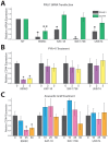
The panel of ATC cell lines was screened for the effect of SUMO inhibition on CD44 RNA expression, determined by RT-PCR. (A) Cell lines indicated were transfected with PIAS1 vs. non-targeting (NT) siRNA and examined for effect on CD44 expression; only 8505C demonstrated significant reduction of CD44 expression. (B/C) The panel of ATC cell lines was tested for a dose-dependent effect on CD44 expression with treatment with PYR-41 with vehicle only (0), 2 μM (2) or 5 μM (5) (panel B) or anacardic acid with vehicle (0), 10 μM (10), 25 μM (25) or 50 μM (50) (panel C) with treatment for 72 hours. Response to SUMO inhibitors was only noted in 8505C.
Figure 5
Figure 5. Repression of CD44 with SUMO Inhibitors in 8505C Cells
The 8505C ATC cell line was tested for a response to SUMO inhibitors by western blot. (A) Cells were treated with PYR-41 at vehicle only (0), 2 μM (2) or 5 μM (5) as indicated. Western blot (top panel) showed reduction of CD44 expression with loss of SUMO-conjugated TFAP2A. FACS analysis (bottom panel) confirmed dose-dependent reduction of cell surface expression of CD44; numbers in colored boxes reports percent CD44-positive cells at each drug concentration and corresponds to flow diagram of the same color. (B) Experiments parallel to A demonstrate dose-dependent reduction of CD44 with AA at vehicle only (0), 10 μM (10), 25 μM (25) or 50 μM (50) as indicated by western blot (top panel) showing repression of CD44 and loss of SUMO-conjugated TFAP2A; bottom panel shows similar dose-dependent repression of cell surface expression of CD44 with AA; numbers in colored boxes reports percent CD44-positive cells at each drug concentration.
Figure 6
Figure 6. Effect of SUMO Inhibition are Mediated by TFAP2A
(A) Western blot of 8505C cells transfected with non-targeting (NT), TFAP2A (A), PIAS1 (P) or combination of TFAP2A and PIAS1 (A+P) siRNA and analyzed for expression of CD44, TFAP2A, PIAS1 or GAPDH as indicated. Relative CD44 protein was NT: 1.0; A: 0.52; P: 0.34; A+P: 0.94. Relative TFAP2A protein was NT: 1.0; A: 0.003; P: 0.51; A+P: 0.02. Relative PIAS1 protein was NT: 1.0; A: 0.93; P: 0.12; A+P: 0.59. (B) Western blot of CD44 expression with vehicle (0) or anacardic acid (AA) at 50 μM (50) transfected with either non-targeting (NT) or TFAP2A (A) siRNA. Relative CD44 protein was NT/0: 1.0; NT/50: 0.13; AA/0: 1.0; AA/50: 0.44. Relative TFAP2A protein was NT/0: 1.0; NT/50: 0.35; A/0: <0.01; A/50: <0.01.
Figure 7
Figure 7. Repression of PIAS1 with Anacardic Acid Treatment
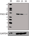
Western blots of protein from 8505C cells treated with vehicle only (VEH) or AA at 25 μM (25) or 50 μM (50) for 96 hours and analyzed for PIAS1 expression with GAPDH loading control; M shows lane with markers. Data demonstrate a dose-dependent repression of PIAS1.
Figure 8
Figure 8. Tumor-free Survival (TFS) of Mice with SUMO Inhibitors
Xenografts were inoculated into mice (n=5 per group) and treated with vehicle (control) or PYR-41 (A) or anacardic acid (B) and examined for tumor growth. Data demonstrates delay in TFS with SUMO inhibitors.
Figure 9
Figure 9. Xenografts of 8505C Analyzed for Growth, H&E and CD44
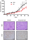
Mice with 8505C xenografts were gavaged with vehicle (VEH) or anacardic acid (AA) and evaluated for total volume of xenografts show a significant reduction in growth rate with AA treatment. *<0.05, **<0.001. Bottom panels show H&E (x200) and immunohistochemistry for CD44 of tumors from vehicle and AA treated animals, as indicated.
Figure 10
Figure 10. Immunohistochemistry for TFAP2A Expression in ATC Tumors
Eleven archival ATC tumor specimens were examined for TFAP2A expression by immunohistochemistry. Tumors 1, 2, 4, and 9 (denoted in green) demonstrated positive brown nuclear TFAP2A staining.
Figure 11
Figure 11. Immunohistochemistry for PIAS1 Expression in ATC Tumors
Eleven archival ATC tumor specimens (same as in Figure 10) were examined for PIAS1 expression by immunohistochemistry. Tumors 1, 2, 3, 4, 7, 9 10 and 11 (denoted in green) demonstrated positive brown PIAS1 staining.
Figure 12
Figure 12. Model of Regulation of CSC/TIC Phenotype Through Sumoylation of TFAP2A
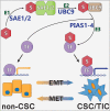
Shown if the proposed model of SUMO conjugation of TFAP2A (TF) controlling patterns of gene expression. SUMO unconjugated TFAP2A is sumoylated through an enzymatic cascade involving E1 SUMO enzymes SAE1/2, E2 SUMO enzyme UBC9 and E3 SUMO ligases (including the PIAS family, PIAS1-4) resulting in SUMO conjugation of target TFAP2A (TF). SUMO unconjugated TFAP2A regulates expression of genes associated with the differentiated non-CSC subtype. SUMO conjugated TFAP2A regulates the pattern of gene expression associated with the CSC/TIC subtype. The model demonstrates the plasticity of CSC/TIC altered through SUMO conjugation of the TFAP2A transcription factor.

References

    1. Hanahan D, Weinberg RA. The hallmarks of cancer. Cell. 2000;100:57–70. - PubMed
    1. Block KI, Gyllenhaal C, Lowe L, Amedei A, Amin AR, Amin A, Aquilano K, Arbiser J, Arreola A, Arzumanyan A, Ashraf SS, Azmi AS, Benencia F, et al. Designing a broad-spectrum integrative approach for cancer prevention and treatment. Semin Cancer Biol. 2015;35:S276–304. - PMC - PubMed
    1. Charafe-Jauffret E, Ginestier C, Bertucci F, Cabaud O, Wicinski J, Finetti P, Josselin E, Adelaide J, Nguyen TT, Monville F, Jacquemier J, Thomassin-Piana J, Pinna G, et al. ALDH1-positive cancer stem cells predict engraftment of primary breast tumors and are governed by a common stem cell program. Cancer Res. 2013;73:7290–300. - PubMed
    1. Dontu G, Al-Hajj M, Abdallah WM, Clarke MF, Wicha MS. Stem cells in normal breast development and breast cancer. Cell Prolif. 2003;36(Suppl 1):59–72. - PMC - PubMed
    1. Iqbal J, Chong PY, Tan PH. Breast cancer stem cells: an update. J Clin Pathol. 2013;66:485–90. - PubMed