

Apparent diffusion coefficient value for a B-cell central nervous system lymphoma in a cat
- PMID: 29383265
- PMCID: PMC5784466
- DOI: 10.1177/2055116917750762
Apparent diffusion coefficient value for a B-cell central nervous system lymphoma in a cat
Abstract
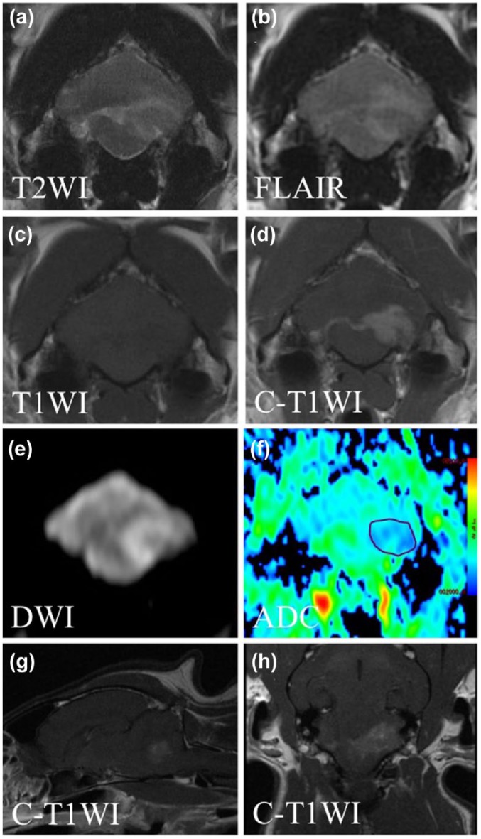
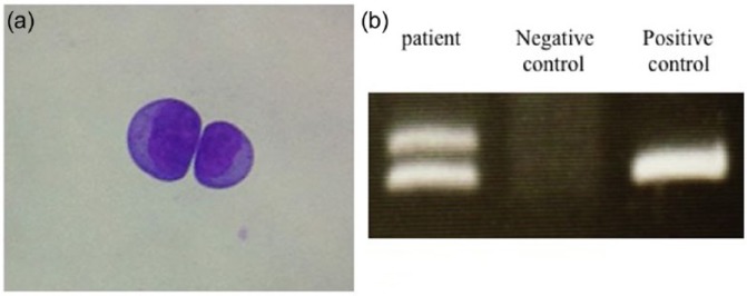
Case summary: This report involves a 10-year-old male mixed-breed cat with a B-cell central nervous system (CNS) lymphoma. The cat presented with ataxia progressing to left hemiparesis. While haematological findings were normal, serum biochemistry showed a high creatine phosphokinase concentration. MRI revealed a homogeneously enhancing well-demarcated extra-axial lesion involving the region of the left lateral aperture with oedema in left flocculus and left medulla oblongata. On diffusion-weighted imaging, the lesion margins showed marked hyperintensity relative to the right cerebellar hemisphere. On an apparent diffusion coefficient map, the lesion appeared hypointense, with an apparent diffusion coefficient value of 0.57 ± 0.01 × 10⁻³ mm2/s. Cerebrospinal fluid (CSF) analysis and cytology, and genetic analysis of CSF lymphoblasts confirmed a diagnosis of B-cell lymphoma. The owner opted for palliative treatment with prednisolone (1 mg/kg q12h); however, the cat died of dyspnoea 10 days after presentation.
Relevance and novel information: CNS lymphomas, which are the second most common intracranial tumours in cats, are highly infiltrative lesions and radical surgical excision is not recommended. Therefore, accurate diagnosis is crucial. However, contrast-enhanced MRI cannot always differentiate these lesions from other conditions, including other CNS tumours and strokes. To the best of our knowledge, this is the first report to document the diffusion-weighted imaging features and apparent diffusion coefficient value for a feline CNS lymphoma. These findings are expected to improve the diagnostic accuracy of these lesions in cats.
Conflict of interest statement
Conflict of interest: The authors declared no potential conflicts of interest with respect to the research, authorship, and/or publication of this article.
Figures
Similar articles
-
MRI findings, including diffusion-weighted imaging and apparent diffusion coefficient value, in two cats with nasopharyngeal polyps and one cat with lymphoma.JFMS Open Rep. 2018 Nov 27;4(2):2055116918812254. doi: 10.1177/2055116918812254. eCollection 2018 Jul-Dec. JFMS Open Rep. 2018. PMID: 30505455 Free PMC article.
-
Clinical and magnetic resonance imaging features of lymphoma involving the nervous system in cats.J Vet Intern Med. 2022 Mar;36(2):679-693. doi: 10.1111/jvim.16350. Epub 2022 Jan 20. J Vet Intern Med. 2022. PMID: 35048412 Free PMC article.
-
Case report: MRI findings with CNS blastomycosis in three domestic cats.Front Vet Sci. 2022 Aug 16;9:966853. doi: 10.3389/fvets.2022.966853. eCollection 2022. Front Vet Sci. 2022. PMID: 36051537 Free PMC article.
-
[A case of progressive multifocal leukoencephalopathy presenting white matter MRI lesions extending over the cerebral cortex and a marked decrease in cerebral blood flow on SPECT, and associated with HTLV-I infection].Rinsho Shinkeigaku. 2005 Jun;45(6):426-30. Rinsho Shinkeigaku. 2005. PMID: 16022467 Review. Japanese.
-
Primary marginal zone B-cell lymphoma of the cavernous sinus: a case report and review of the literature.BMC Med Imaging. 2021 Feb 12;21(1):25. doi: 10.1186/s12880-021-00556-w. BMC Med Imaging. 2021. PMID: 33579209 Free PMC article. Review.
Cited by
-
Feline lymphoma of the nervous system. Immunophenotype and anatomical patterns in 24 cases.Front Vet Sci. 2022 Sep 8;9:959466. doi: 10.3389/fvets.2022.959466. eCollection 2022. Front Vet Sci. 2022. PMID: 36157173 Free PMC article.
-
MRI findings, including diffusion-weighted imaging, in seven cats with nasal lymphoma and two cats with nasal adenocarcinoma.J Feline Med Surg. 2021 Apr;23(4):393-399. doi: 10.1177/1098612X20932819. Epub 2020 Jun 23. J Feline Med Surg. 2021. PMID: 32573314 Free PMC article.
-
Neuropathology of Central and Peripheral Nervous System Lymphoma in Dogs and Cats: A Study of 92 Cases and Review of the Literature.Animals (Basel). 2023 Feb 27;13(5):862. doi: 10.3390/ani13050862. Animals (Basel). 2023. PMID: 36899719 Free PMC article.
-
Successful Treatment of Central Nervous System Lymphoma with Combination Therapy of Nimustine and Prednisolone in Two Dogs.Vet Sci. 2023 Aug 22;10(9):533. doi: 10.3390/vetsci10090533. Vet Sci. 2023. PMID: 37756055 Free PMC article.
-
MRI findings, including diffusion-weighted imaging and apparent diffusion coefficient value, in two cats with nasopharyngeal polyps and one cat with lymphoma.JFMS Open Rep. 2018 Nov 27;4(2):2055116918812254. doi: 10.1177/2055116918812254. eCollection 2018 Jul-Dec. JFMS Open Rep. 2018. PMID: 30505455 Free PMC article.
References
Publication types
LinkOut - more resources
Full Text Sources
Other Literature Sources
Miscellaneous