. 2018 Oct;46(5):409-418.
doi: 10.1007/s00240-018-1035-0. Epub 2018 Jan 30.

Calcium receptor signaling and citrate transport

Affiliations

Calcium receptor signaling and citrate transport

Ryan W Walker et al. Urolithiasis. 2018 Oct.

Abstract

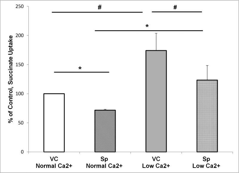
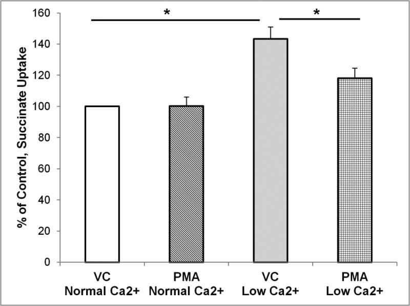
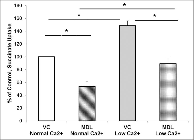
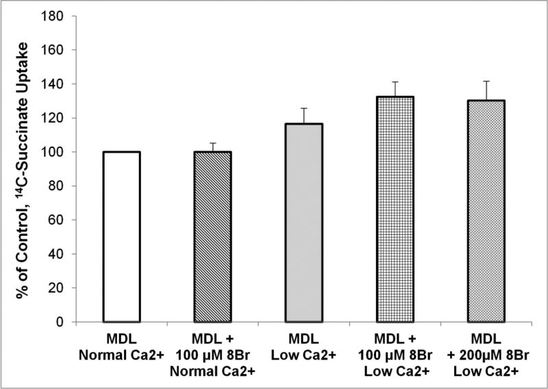
The calcium sensing receptor (CaSR) in the distal nephron decreases the propensity for calcium stones. Here we investigate if the apical CaSR in the proximal tubule also prevents stone formation acting via regulation of apical dicarboxylate and citrate transport. Urinary citrate, partially reabsorbed as a dicarboxylate in the proximal tubule lumen, inhibits stone formation by complexing calcium. We previously demonstrated a novel apical calcium-sensitive dicarboxylate transport system in OK proximal tubule cells. This calcium-sensitive process has the potential to modulate the amount of citrate available to complex increased urinary calcium. Using isotope labeled succinate uptake in OK cells along with various pharmacologic tools we examined whether the CaSR alters apical dicarboxylate transport and through which signal transduction pathways this occurs. Our results indicate that in the proximal tubule CaSR adjusts apical dicarboxylate transport, and does so via a CaSR → Gq → PKC signaling pathway. Thus, the CaSR may decrease the propensity for stone formation via actions in both proximal and distal nephron segments.

Keywords: Apical; CaSR; Calcium-sensitive; Citrate; Dicarboxylate transport; Proximal tubule.


Conflict of interest statement

Conflict of Interest: Dr Ryan Walker declares that he has no conflict of interest. Dr Shijia Zhang declares that he has no conflict of interest. Ms. Joycelynn Coleman-Barnett declares that she has no conflict of interest. Dr L. Lee Hamm declares that he has no conflict of interest. Dr Kathleen Hering-Smith declares that she has no conflict of interest.

Figures

Figure 1
Figure 1. Proximal Tubule Apical CaSR Signaling Cascade
The apical proximal tubule CaSR potentially signals via the G-proteins: Gq, Gi or Gs in OK cell monolayers. Figure 1 illustrates an overview of CaSR signaling, the G-protein signaling pathways studied and pharmacologicals used to determine the role of extracellular Ca2+ in apical dicarboxylate transport regulation in OK proximal tubule cell monolayers. Time of treatment, concentration and vehicle controls are outlined in Table 1. Green arrows indicate stimulation, while red bars indicate inhibition by the pharmacological agents used in this study. Black arrows, ↑, represent stimulation or increases and bars, ⊢, inhibition of some of the major components of G-protein coupled signaling cascade.
Figure 2
Figure 2. Apical CaSR activation results in inhibition of apical dicarboxylate transport
We have shown in our previously published studies () and herein that decreasing extracellular Ca2+, [Ca2+]o, results in augmentation of apical dicarboxylate transport (compare Bar 3 from left with Bar 1) in the OK proximal tubule cell line. Succinate transport increased to 174 ± 29.8% of control in low [Ca2+]o compared to normal [Ca2+]o. Addition of spermine (1 mM, Sp) to activate the apical CaSR resulted in decreasing succinate transport in both normal (Bar 2) and low [Ca2+]o (Bar 4). Spermine addition decreased succinate transport in normal [Ca2+]o (Bar 2) to 71.6 ± 1.7% of control, p<0.05. In low [Ca2+]o (Bar 3, VC) succinate transport was 174 ± 29.8% of that in normal [Ca2+]o; addition of spermine to low [Ca2+]o reduced dicarboxylate transport to 123 ± 25.3% of control, p<0.01 (Bar 4). Taken together the addition of spermine to OK cell monolayers results in decreased dicarboxylate transport indicating the CaSR is involved in the regulation of apical dicarboxylate transport in the proximal tubule. # = p<0.05, * = p<0.01 VC = vehicle control.
Figure 3
Figure 3. Gq signaling is involved in dicarboxylate transport regulation in both normal and low [Ca2+]o
Activation of apical CaSR may result in signaling via one of 3 G-proteins as illustrated in Figure 1. This series of experiments looks at Gq signaling and used thapsigargin (Thaps) pretreatment (10 µM, 30 min) to increase intracellular calcium [Ca2+]i and thus mimic signaling via Gq. Thaps pretreatment resulted in decreased dicarboxylate transport in both normal (100 ± 0.0 vs 73.5 ± 2.9% of control, p<0.01) and low [Ca2+]o (134 ± 7.2 vs 100 ± 8.2% of control, p<0.01). * = p<0.01. VC = vehicle control. This indicates that following CaSR activation by calcium, Gq signaling is involved in apical dicarboxylate transport regulation.
Figure 4
Figure 4. PKC Activation
Gq signaling produces increases in intracellular Ca2+ and is known to activate PKC. Since increasing [Ca2+]i with Thaps shown in Figure 3 is involved, our next step was to directly activate PKC and examine changes in transport. OK cell monolayers were pretreated with Phorbol 12-myristate 13-acetate (50 nM PMA) for 30 min to activate endogenous PKC. Succinate transport in normal [Ca2+]o was unchanged (100 ± 0.0 vs 100 ± 5.9% of control, p=NS). However, PMA addition resulted in decreased dicarboxylate transport in low [Ca2+]o (143 ± 7.8 vs 118 ± 6.6% of control, p<0.01). Thus when changes/decreases in [Ca2+]o occur, CaSR signaling via Gq →PKC is involved in apical dicarboxylate transport regulation. Thus PKC activation is likely part of the signaling cascade (Figure 1) that results in lower levels of apical dicarboxylate transport in normal concentrations of [Ca2+]o. VC = vehicle control. * = p<0.01.
Figure 5
Figure 5. Direct inhibition of Gi protein with PTX does not alter dicarboxylate transport
Possible CaSR signaling via the Gi protein can be investigated by using pertussis toxin (PTX) to directly inhibit Gi. Signaling via Gi (Figure 1) results in the inhibition of adenylate cyclase (AC). PTX, used to block the inhibition of AC, should result in an increase of intracellular cAMP. OK cell monolayers were treated overnight with PTX (100 ng/ml) to inhibit Gi. Shown here PTX pretreatment did not alter dicarboxylate transport in either normal (100 ± 0.0 vs 107 ± 1.9% of control, p=NS) or low [Ca2+]o (174 ± 24 vs 178 ± 15% of control, p=NS). VC = vehicle control. * = p<0.01.
Figure 6
Figure 6. Direct inhibition of AC with MDL decreased dicarboxylate transport in both normal and low [Ca2+]o
Another way to investigate Gi signaling is to directly inhibit AC by the addition of MDL-12,330A (MDL). OK cell monolayers were treated with 50 µM of MDL for 30 min before beginning transport measurements. MDL inhibited succinate transport in both normal (100 ± 0.00 vs 53.7 ± 7.4% of control, p<0.01) and low [Ca2+]o (146 ± 6.4 vs 88.7 ± 9.0% of control, p<0.01). VC = vehicle control. * = p<0.01. The MDL results differ from the results in Figure 5, where PTX had no effect on dicarboxylate transport; this discrepancy is addressed in the text.
Figure 7
Figure 7. Increasing intracellular cAMP does not offset the effects of AC inhibition on dicarboxylate transport
In this series of experiments MDL was used as a direct AC inhibitor to block increases in endogenously generated intracellular cAMP in all 4 groups of experiments. 8-Br-cAMP (8Br, 100 µM) is a cell membrane permeable form of cAMP (8) and was used to increase intracellular cAMP exogenously. Comparing Bars 1 vs 2, increasing intracellular cAMP by the addition of 8Br did not overcome the direct inhibition of AC by MDL in normal [Ca2+]o (100 ± 0.00 vs 100 ± 5.1% of control). Similar results were found in low [Ca2+]o; 8Br had no significant effect in the presence of MDL in low [Ca2+]o (116 ± 9.2 vs 133 ± 8.7% of control) shown in Bars 3 vs 4. And when 8Br concentrations were doubled to 200 µM, again there was no significant effect (116 ± 9.2 vs 130 ± 11.3% of control) shown in Bars 3 vs 5. This provides further evidence that inhibition of dicarboxylate transport in both normal and low [Ca2+]o (Figure 6) is not via AC inhibition, indicating that MDL may be acting via another mechanism.
Figure 8
Figure 8. Increasing intracellular cAMP via 8-Br-cAMP has no effect on dicarboxylate transport
Intracellular cAMP in OK cell monolayers was increased by the addition of 100 µM 8-Br-cAMP (8Br) for 30 min before start of transport measurements. Raising intracellular cAMP had no effect on dicarboxylate transport in either normal (100 ± 0.00 vs 94.7 ± 1.7% of control, p=NS) or low (154 ± 9.9 vs 131 ± 9.6% of control, p=NS) [Ca2+]o. VC = vehicle control. * = p<0.01. This suggests that CaSR activation is not regulating dicarboxylate transport via Gs. Importantly, these results align with those in Figure 5 and 7, indicating that neither Gi nor Gs are involved in regulation of dicarboxylate transport via CaSR activation.
Figure 9
Figure 9. Assay of Intracellular cAMP Levels
To further examine if there was a role of cAMP in dicarboxylate transport regulation, intracellular levels of cAMP were measured. Shown here, no changes in intracellular cAMP levels were found in low [Ca2+]o (0.335 ± 0.103 vs 0.272 ± 0.095 pmol/ml, p=NS). The CaSR activator spermine (1 mM, 5 min) also had no significant effect on intracellular cAMP levels in normal [Ca2+]o compared to normal control (0.335 ± 0.103 vs 0.354 ± 0.136 pmol/ml, p=NS). This indicates that CaSR activation in OK cell monolayers does not result in AC activation or signal via Gi or Gs which act via changes in intracellular cAMP. As a positive control for the cAMP assays, the AC activator forskolin (10 µM, 30 min) significantly increased endogenous intracellular cAMP in normal [Ca2+]o (0.335 ± 0.103 vs 1.008 ± 0.228 pmol/ml, p<0.05). These results further indicate that activation of the CaSR to regulate dicarboxylate transport in OK cells is not signaling via Gi or Gs. # = p<0.05.

Similar articles

Cited by

References

    1. Egbuna O, Quinn S, Kantham L, Butters R, Pang J, Pollak M, Goltzman D, Brown E. The full-length calcium-sensing receptor dampens the calcemic response to 1alpha,25(OH)2 vitamin D3 in vivo independently of parathyroid hormone. Am J Physiol Renal Physiol. 2009;297(3):F720–728. - PMC - PubMed
    1. Quamme GA. Effect of hypercalcemia on renal tubular handling of calcium and magnesium. Can J Physiol Pharmacol. 1982;60(10):1275–1280. - PubMed
    1. Riccardi D, Brown EM. Physiology and pathophysiology of the calcium-sensing receptor in the kidney. Am J Physiol Renal Physiol. 2010;298(3):F485–499. - PMC - PubMed
    1. Wang WH, Lu M, Hebert SC. Cytochrome P-450 metabolites mediate extracellular Ca(2+)-induced inhibition of apical K+ channels in the TAL. Am J Physiol. 1996;271(1 Pt 1):C103–111. - PubMed
    1. Topala CN, Schoeber JP, Searchfield LE, Riccardi D, Hoenderop JG, Bindels RJ. Activation of the Ca2+-sensing receptor stimulates the activity of the epithelial Ca2+ channel TRPV5. Cell Calcium. 2009;45(4):331–339. - PubMed

LinkOut - more resources