

Normalization of ADC does not improve correlation with overall survival in patients with high-grade glioma (HGG)
- PMID: 29383647
- PMCID: PMC6071871
- DOI: 10.1007/s11060-017-2719-y
Normalization of ADC does not improve correlation with overall survival in patients with high-grade glioma (HGG)
Abstract
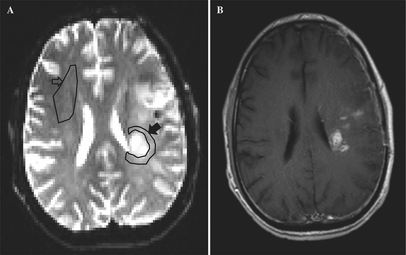
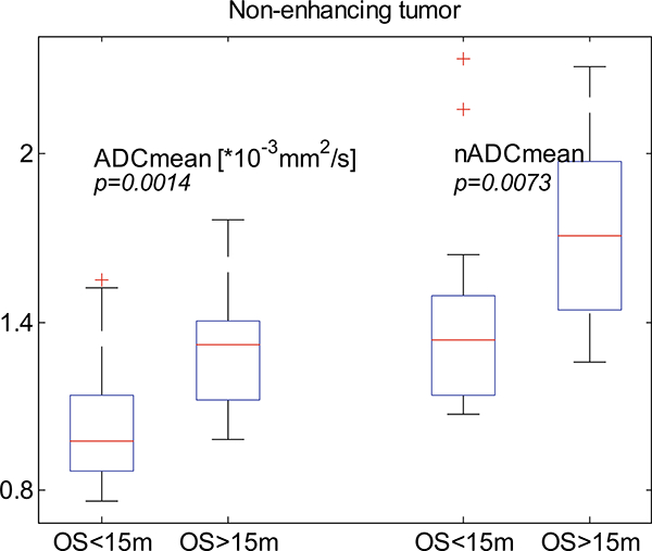
Mixed reports leave uncertainty about whether normalization of apparent diffusion coefficient (ADC) to a within-subject white matter reference is necessary for assessment of tumor cellularity. We tested whether normalization improves the previously reported correlation of resection margin ADC with 15-month overall survival (OS) in HGG patients. Spin-echo echo-planar DWI was retrieved from 3 T MRI acquired between maximal resection and radiation in 37 adults with new-onset HGG (25 glioblastoma; 12 anaplastic astrocytoma). ADC maps were produced with the FSL DTIFIT tool (Oxford Centre for Functional MRI). 3 neuroradiologists manually selected regions of interest (ROI) in normal appearing white matter (NAWM) and in non-enhancing tumor (NT) < 2 cm from the margin of residual enhancing tumor or resection cavity. Normalized ADC (nADC) was computed as the ratio of absolute NT ADC to NAWM ADC. Reproducibility of nADC and absolute ADC among the readers' ROI was assessed using intra-class correlation coefficient (ICC) and within-subject coefficient of variation (wCV). Correlations of ADC and nADC with OS were compared using receiver operating characteristics (ROC) analysis. A p value 0.05 was considered statistically significant. Both mean ADC and nADC differed significantly between patients subgrouped by 15-month OS (p = 0.0014 and 0.0073 respectively). wCV and ICC among the readers were similar for absolute and normalized ADC. In ROC analysis of correlation with OS, nADC did not perform significantly better than absolute ADC. Normalization does not significantly improve the correlation of absolute ADC with OS in HGG, suggesting that normalization is not necessary for clinical or research ADC analysis in HGG patients.
Keywords: ADC normalization; Apparent diffusion coefficient (ADC); Diffusion weighted imaging (DWI); High-grade glioma (HGG).
Conflict of interest statement
Compliance with ethical standards
Figures

References
-
- Wen PY, Macdonald DR, Reardon DA, Cloughesy TF, Sorensen AG, Galanis E, Degroot J, Wick W, Gilbert MR, Lassman AB, Tsien C, Mikkelsen T, Wong ET, Chamberlain MC, Stupp R, Lamborn KR, Vogelbaum MA, van den Bent MJ, Chang SM (2010) Updated response assessment criteria for high-grade gliomas: response assessment in neuro-oncology working group. J Clin Oncol 28(11):1963–1972. 10.1200/jco.2009.26.3541 - DOI - PubMed
-
- Padhani AR, Liu G, Mu-Koh D, Chenevert TL, Thoeny HC, Takahara T, Dzik-Jurasz A, Ross BD, Van Cauteren M, Collins D, Hammoud DA, Rustin GJS, Taouli B, Choyke PL (2009) Diffusion-weighted magnetic resonance imaging as a cancer biomarker: consensus and recommendations. Neoplasia 11(2):102–125 - PMC - PubMed
-
- Murakami R, Sugahara T, Nakamura H, Hirai T, Kitajima M, Hayashida Y, Baba Y, Oya N, Kuratsu J, Yamashita Y (2007) Malignant supratentorial astrocytoma treated with postoperative radiation therapy: prognostic value of pretreatment quantitative diffusion-weighted MR imaging. Radiology 243(2):493–499. 10.1148/radiol.2432060450 - DOI - PubMed
-
- Yamasaki F, Sugiyama K, Ohtaki M, Takeshima Y, Abe N, Akiyama Y, Takaba J, Amatya VJ, Saito T, Kajiwara Y, Hanaya R, Kurisu K (2010) Glioblastoma treated with postoperative radiochemotherapy: prognostic value of apparent diffusion coefficient at MR imaging. Eur J Radiol 73(3):532–537. 10.1016/j.ejrad.2009.01.013 - DOI - PubMed
MeSH terms
Grants and funding
LinkOut - more resources
Full Text Sources
Other Literature Sources
Medical
Research Materials
