

Neutrophilia, gelatinase release and microvascular leakage induced by human mast cell tryptase in a mouse model: Lack of a role of protease-activated receptor 2 (PAR2)
- PMID: 29383785
- PMCID: PMC5969079
- DOI: 10.1111/cea.13108
Neutrophilia, gelatinase release and microvascular leakage induced by human mast cell tryptase in a mouse model: Lack of a role of protease-activated receptor 2 (PAR2)
Abstract
Background: Tryptase, the most abundant protease of the human mast cell, has been implicated as a key mediator of allergic inflammation that acts through activation of PAR2.
Objectives: To investigate the contribution of PAR2 in the pro-inflammatory actions mediated by tryptase in a mice model.
Methods: We have injected recombinant human βII-tryptase into the peritoneum of PAR2-deficient and wild-type C57BL/6 mice. After 6, 12 and 24 hours, mice were killed, peritoneal lavage performed and inflammatory changes investigated.
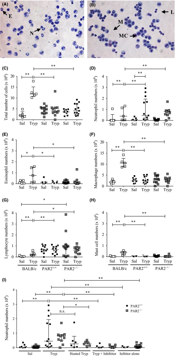
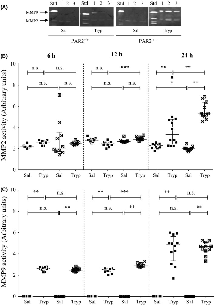
Results: Tryptase stimulated an increase in neutrophil numbers in the peritoneum, but responses did not differ between PAR2-deficient and wild-type mice. Heat inactivation of tryptase or pre-incubation with a selective tryptase inhibitor reduced neutrophilia, but neutrophil accumulation was not elicited with a peptide agonist of PAR2 (SLIGRL-NH2 ). Zymography indicated that tryptase stimulated the release of matrix metalloproteinases (MMP) 2 and 9 in the peritoneum of both mouse strains. Studies involving immunomagnetic isolation of neutrophils suggested that neutrophils represent the major cellular source of tryptase-induced MMP2 and MMP9. At 24 hours after tryptase injection, there was increased microvascular leakage as indicated by high levels of albumin in peritoneal lavage fluid, and this appeared to be partially abolished by heat-inactivating tryptase or addition of a protease inhibitor. There was no corresponding increase in levels of histamine or total protein. The extent of tryptase-induced microvascular leakage or gelatinase release into the peritoneum did not differ between PAR2-deficient and wild-type mice.
Conclusions: Our findings indicate that tryptase is a potent stimulus for neutrophil accumulation, MMP release and microvascular leakage. Although these actions required an intact catalytic site, the primary mechanism of tryptase in vivo would appear to involve processes independent of PAR2.
Keywords: allergy; inflammation; mast cells/basophils; neutrophils; transgenic/knockout mice.
© 2018 The Authors. Clinical & Experimental Allergy Published by John Wiley & Sons Ltd.
Figures

Similar articles
-
A role for mast cells and mast cell tryptase in driving neutrophil recruitment in LPS-induced lung inflammation via protease-activated receptor 2 in mice.Inflamm Res. 2020 Oct;69(10):1059-1070. doi: 10.1007/s00011-020-01376-4. Epub 2020 Jul 6. Inflamm Res. 2020. PMID: 32632517
-
Human mast cell tryptase: a stimulus of microvascular leakage and mast cell activation.Eur J Pharmacol. 1997 Jun 5;328(1):89-97. doi: 10.1016/s0014-2999(97)83033-6. Eur J Pharmacol. 1997. PMID: 9203574
-
Protease activated-receptor 2 is necessary for neutrophil chemorepulsion induced by trypsin, tryptase, or dipeptidyl peptidase IV.J Leukoc Biol. 2018 Jan;103(1):119-128. doi: 10.1002/JLB.3A0717-308R. Epub 2017 Dec 11. J Leukoc Biol. 2018. PMID: 29345066 Free PMC article.
-
Mast cell tryptases in allergic inflammation and immediate hypersensitivity.Curr Opin Immunol. 2021 Oct;72:94-106. doi: 10.1016/j.coi.2021.04.001. Epub 2021 Apr 28. Curr Opin Immunol. 2021. PMID: 33932709 Review.
-
Tryptase, a novel link between allergic inflammation and fibrosis.Trends Immunol. 2003 Apr;24(4):158-61. doi: 10.1016/s1471-4906(03)00058-9. Trends Immunol. 2003. PMID: 12697439 Review.
Cited by
-
Biomarkers and Their Possible Functions in the Intestinal Microenvironment of Chagasic Megacolon: An Overview of the (Neuro)inflammatory Process.J Immunol Res. 2021 Apr 7;2021:6668739. doi: 10.1155/2021/6668739. eCollection 2021. J Immunol Res. 2021. PMID: 33928170 Free PMC article. Review.
-
Effect of tryptase on mouse brain microvascular endothelial cells via protease-activated receptor 2.J Neuroinflammation. 2018 Aug 31;15(1):248. doi: 10.1186/s12974-018-1287-1. J Neuroinflammation. 2018. PMID: 30170602 Free PMC article.
-
Pruritogens in pemphigoid diseases: Possible therapeutic targets for a burdensome symptom.J Dermatol. 2023 Feb;50(2):150-161. doi: 10.1111/1346-8138.16652. Epub 2022 Dec 7. J Dermatol. 2023. PMID: 36477831 Free PMC article. Review.
-
Glia Maturation Factor and Mast Cell-Dependent Expression of Inflammatory Mediators and Proteinase Activated Receptor-2 in Neuroinflammation.J Alzheimers Dis. 2018;66(3):1117-1129. doi: 10.3233/JAD-180786. J Alzheimers Dis. 2018. PMID: 30372685 Free PMC article.
-
Expression of Inflammatory Markers RANK, MMP-9 and PTHrP in Chronic Apical Periodontitis from People Living with HIV Undergoing Antiretroviral Therapy.J Clin Med. 2020 Nov 9;9(11):3611. doi: 10.3390/jcm9113611. J Clin Med. 2020. PMID: 33182451 Free PMC article.
References
-
- Molino M, Barnathan ES, Numerof R, et al. Interactions of mast cell tryptase with thrombin receptors and PAR‐2. J Biol Chem. 1997;272:4043‐4049. - PubMed
-
- Mirza H, Schmidt VA, Derian CK, Jesty J, Bahou WF. Mitogenic responses mediated through the proteinase‐activated receptor‐2 are induced by expressed forms of mast cell alpha‐ or beta‐tryptases. Blood. 1997;90:3914‐3922. - PubMed
-
- Wakita H, Furukawa F, Takigawa M. Thrombin and trypsin induce granulocyte‐macrophage colony‐stimulating factor and interleukin‐6 gene expression in cultured normal human keratinocytes. Proc Assoc Am Physicians. 1997;109:190‐207. - PubMed
-
- Vliagoftis H, Befus AD, Hollenberg MD, Moqbel R. Airway epithelial cells release eosinophil survival‐promoting factors (GM‐CSF) after stimulation of proteinase‐activated receptor 2. J Allergy Clin Immunol. 2001;107:679‐685. - PubMed
Publication types
MeSH terms
Substances
Grants and funding
LinkOut - more resources
Full Text Sources
Other Literature Sources
Medical
Miscellaneous
