

Comparing human and mouse salivary glands: A practice guide for salivary researchers
- PMID: 29383862
- PMCID: PMC6613660
- DOI: 10.1111/odi.12840
Comparing human and mouse salivary glands: A practice guide for salivary researchers
Abstract
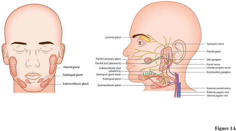
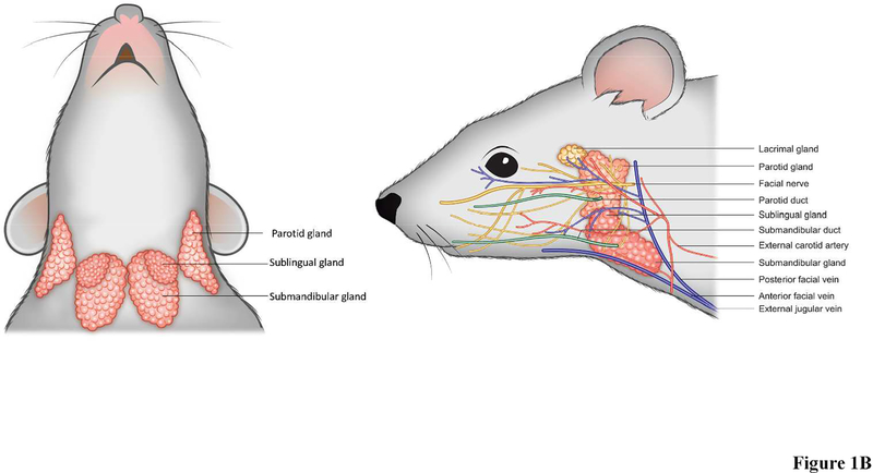
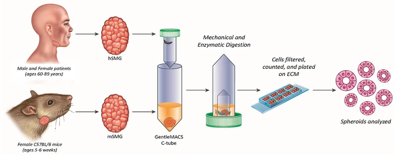
Mice are a widely utilized in vivo model for translational salivary gland research but must be used with caution. Specifically, mouse salivary glands are similar in many ways to human salivary glands (i.e., in terms of their anatomy, histology, and physiology) and are both readily available and relatively easy and affordable to maintain. However, there are some significant differences between the two organisms, and by extension, the salivary glands derived from them must be taken into account for translational studies. The current review details pertinent similarities and differences between human and mouse salivary glands and offers practical guidelines for using both for research purposes.
Keywords: anatomy; histology; physiology; primary cells; tissue culture; tissue engineering.
© 2018 John Wiley & Sons A/S. Published by John Wiley & Sons Ltd. All rights reserved.
Figures
Similar articles
-
Anatomy, biogenesis and regeneration of salivary glands.Monogr Oral Sci. 2014;24:1-13. doi: 10.1159/000358776. Epub 2014 May 23. Monogr Oral Sci. 2014. PMID: 24862590 Free PMC article. Review.
-
Physiology, Pathology and Regeneration of Salivary Glands.Cells. 2019 Aug 26;8(9):976. doi: 10.3390/cells8090976. Cells. 2019. PMID: 31455013 Free PMC article. Review.
-
[Pathology of the salivary glands. I: Introduction to the anatomy and physiology of the salivary gland. Approach to the patient with salivary gland disease].Dent Cadmos. 1984 Dec;52(12):151-2, 154-5. Dent Cadmos. 1984. PMID: 6598784 Italian. No abstract available.
-
Histochemical comparison of the resting and exhausted cells of the pancreas and the salivary glands of the mouse and rat.Anat Rec. 1947 Mar;97(3):359. Anat Rec. 1947. PMID: 20289373 No abstract available.
-
Cross-Sectional Imaging Techniques and Normal Anatomy of the Salivary Glands.Neuroimaging Clin N Am. 2018 May;28(2):137-158. doi: 10.1016/j.nic.2018.01.001. Epub 2018 Mar 7. Neuroimaging Clin N Am. 2018. PMID: 29622110 Review.
Cited by
-
Strategies for Developing Functional Secretory Epithelia from Porcine Salivary Gland Explant Outgrowth Culture Models.Biomolecules. 2019 Oct 25;9(11):657. doi: 10.3390/biom9110657. Biomolecules. 2019. PMID: 31717706 Free PMC article.
-
Long-Term Regenerative Potential of Submandibular Glands in Albino Rats Following Radiotherapy: Role of Cytokeratin 17 Redistribution.Cureus. 2025 Mar 30;17(3):e81465. doi: 10.7759/cureus.81465. eCollection 2025 Mar. Cureus. 2025. PMID: 40303535 Free PMC article.
-
Predicting Resolvin D1 Pharmacokinetics in Humans with Physiologically-Based Pharmacokinetic Modeling.Clin Transl Sci. 2021 Mar;14(2):683-691. doi: 10.1111/cts.12930. Epub 2020 Nov 30. Clin Transl Sci. 2021. PMID: 33202089 Free PMC article.
-
Protocol for parotidectomy and saliva analysis in mice.STAR Protoc. 2021 Dec 16;3(1):101048. doi: 10.1016/j.xpro.2021.101048. eCollection 2022 Mar 18. STAR Protoc. 2021. PMID: 34977687 Free PMC article.
-
Transduction of Salivary Gland Acinar Cells with a Novel AAV Vector 44.9.Mol Ther Methods Clin Dev. 2020 Oct 14;19:459-466. doi: 10.1016/j.omtm.2020.10.006. eCollection 2020 Dec 11. Mol Ther Methods Clin Dev. 2020. PMID: 33294494 Free PMC article.
References
-
- Alevizos I, Zheng C, Cotrim AP, Liu S, McCullagh L, Billings ME, Goldsmith CM, Tandon M, Helmerhorst EJ, Catalan MA, Danielides SJ, Perez P, Nikolov NP, Chiorini JA, Melvin JE, Oppenheim FG, Illei GG and Baum BJ (2017). Late responses to adenoviral-mediated transfer of the aquaporin-1 gene for radiation-induced salivary hypofunction. Gene Ther 24: 176–186. - PMC - PubMed
-
- Anderson L and Garrett J (1998). Neural regulation of blood flow in the rat submandibular gland. European journal of morphology 36: 213–218. - PubMed
Publication types
MeSH terms
Grants and funding
LinkOut - more resources
Full Text Sources
Other Literature Sources
