Review
. 2018 May 1;119(5):1782-1794.
doi: 10.1152/jn.00331.2017. Epub 2018 Jan 31.

Escape from homeostasis: spinal microcircuits and progression of amyotrophic lateral sclerosis

Affiliations
Review

Escape from homeostasis: spinal microcircuits and progression of amyotrophic lateral sclerosis

Robert M Brownstone et al. J Neurophysiol. .

Abstract

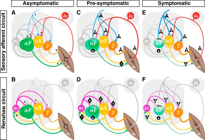
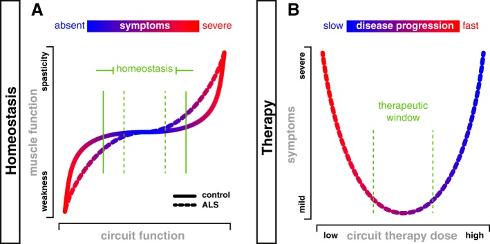
In amyotrophic lateral sclerosis (ALS), loss of motoneuron function leads to weakness and, ultimately, respiratory failure and death. Regardless of the initial pathogenic factors, motoneuron loss follows a specific pattern: the largest α-motoneurons die before smaller α-motoneurons, and γ-motoneurons are spared. In this article, we examine how homeostatic responses to this orderly progression could lead to local microcircuit dysfunction that in turn propagates motoneuron dysfunction and death. We first review motoneuron diversity and the principle of α-γ coactivation and then discuss two specific spinal motoneuron microcircuits: those involving proprioceptive afferents and those involving Renshaw cells. Next, we propose that the overall homeostatic response of the nervous system is aimed at maintaining force output. Thus motoneuron degeneration would lead to an increase in inputs to motoneurons, and, because of the pattern of neuronal degeneration, would result in an imbalance in local microcircuit activity that would overwhelm initial homeostatic responses. We suggest that this activity would ultimately lead to excitotoxicity of motoneurons, which would hasten the progression of disease. Finally, we propose that should this be the case, new therapies targeted toward microcircuit dysfunction could slow the course of ALS.

Keywords: Renshaw cells; excitotoxicity; muscle spindles; proprioceptive afferents; α-motoneurons; γ-motoneurons.


Figures

Fig. 1.
Fig. 1.
Hypothesis: α-motoneuron (α-MN) death leads to microcircuit imbalance and disease progression. Sensory afferent circuits (A, C, E) and Renshaw circuits (B, D, F) in asymptomatic (A, B), presymptomatic (C, D), and symptomatic (E, F) amyotrophic lateral sclerosis (ALS). A and B: normal spinal MN circuits. MN pools comprise multiple types: α-MNs can be defined by the extrafusal muscle fiber (m) types they innervate as either fast (αF; FF and FR types depicted together) or slow (αS) (a). γ-MNs innervate muscle spindles (sp; b), which convey length and velocity information back to α-MNs primarily via group Ia afferents (c), which form monosynaptic connexions with α-MNs (d, e). During most movement, α- and γ-MNs are coactivated by spinal and supraspinal neurons (f). α-MNs also innervate Renshaw cells (RC; g), which in turn inhibit both α- (h) and γ-MNs (i). C and D: in presymptomatic ALS, α-MNs (F-type) become dysfunctional and start to die (*), but γ-MNs are preserved. Homeostatic mechanisms include increased input to α-MNs from spinal and supraspinal circuits (1) to ensure that force production is preserved (2). Thus the input to the coactivated γ-MNs would also increase (3), leading to increased intrafusal fiber contraction (4) out of proportion to extrafusal fibers. This α-γ imbalance would result in an increase in spindle afferent input to α-MNs (6). The increasing glutamatergic (Glu) excitation from these inputs would initially maintain the homeostatic response despite a reduction of activity of fast high-force-producing muscle fibers. In addition, the loss of α-MNs (particularly type F; *) would concomitantly lead to a reduction of output from MN pools to RCs, initially compensated by increased α-MN activity (particularly type S; 7). Thus Renshaw inhibition would at first be maintained in all MN types (8, 9). Together, these processes would lead to increased glutamatergic excitation of vulnerable α-MNs and, hence, excitotoxicity. E and F: in symptomatic stages, type F α-MNs continue to die and type S α-MNs start to degenerate and ultimately die at later stages of the disease (*), but γ-MNs are completely spared. The processes that started in presymptomatic stages would continue, there would be runaway from homeostatic processes, and further excitotoxicity would lead to disease progression. It would no longer be possible to maintain muscle contraction (2), compounding the α-γ imbalance (2 and 4, 5), and the resulting loss of input to RCs (7) would reduce Renshaw inhibition of α-MNs (8) and also diminish γ-MN inhibition (9), thereby contributing to increased excitation of remaining α-MNs but a further imbalance of α-γ output. Note that the thickness of each line represents the integral of synaptic transmission over the number of synaptic contacts on the target cells. In the interest of simplicity and clarity, static and dynamic γ-MNs are represented as a single population, group II sensory afferents are not shown, and MN types are represented as distinct groups, although they are intermingled in each MN pool. Arrows indicate direction of change, and diamonds indicate no net change.
Fig. 2.
Fig. 2.
Homeostasis and microcircuit therapy: targeting microcircuits to slow progression? A: chair-shaped homeostatic curve (Nijhout et al. 2014) demonstrating a region in which normal motor function can be maintained despite increases and decreases in circuit function (solid green vertical bars). Increases beyond this range would lead to positive motor symptoms such as spasticity, whereas reductions would lead to weakness. As circuits degenerate in amyotrophic lateral sclerosis (ALS) and fewer α-motoneurons (α-MNs) are available to these circuits, the homeostatic plateau would narrow (dashed green vertical bars). B: microcircuit therapy for ALS, as defined here, would be aimed at reversing at least one of the arrows in Fig. 1. For example, a therapy to reduce γ-MN activity, or to increase Renshaw cell activity, could reverse the imbalance in these circuits, potentially slowing MN death as depicted by the color scale. We predict that this would reduce symptoms by preserving α-MNs. However, at higher “doses,” such therapy could in itself lead to weakness through reducing α-MN activity. We suggest that there would be a therapeutic window (dashed green vertical bars) in which progression could be slowed and the duration of time that people will have functional muscle contraction would increase, thus leading to improvements in quality of life.

References

    1. Adal MN, Barker D. Intramuscular branching of fusimotor fibres. J Physiol 177: 288–299, 1965. doi:10.1113/jphysiol.1965.sp007592. - DOI - PMC - PubMed
    1. Alvarez FJ, Dewey DE, McMillin P, Fyffe RE. Distribution of cholinergic contacts on Renshaw cells in the rat spinal cord: a light microscopic study. J Physiol 515: 787–797, 1999. doi:10.1111/j.1469-7793.1999.787ab.x. - DOI - PMC - PubMed
    1. Alvarez FJ, Fyffe RE. The continuing case for the Renshaw cell. J Physiol 584: 31–45, 2007. doi:10.1113/jphysiol.2007.136200. - DOI - PMC - PubMed
    1. Appelberg B, Hulliger M, Johansson H, Sojka P. Actions on gamma-motoneurones elicited by electrical stimulation of group I muscle afferent fibres in the hind limb of the cat. J Physiol 335: 237–253, 1983. doi:10.1113/jphysiol.1983.sp014531. - DOI - PMC - PubMed
    1. Arundine M, Tymianski M. Molecular mechanisms of glutamate-dependent neurodegeneration in ischemia and traumatic brain injury. Cell Mol Life Sci 61: 657–668, 2004. doi:10.1007/s00018-003-3319-x. - DOI - PMC - PubMed

Publication types

MeSH terms

LinkOut - more resources