

Forensic age estimation based on development of third molars: a staging technique for magnetic resonance imaging
- PMID: 29384743
- PMCID: PMC6100221
Forensic age estimation based on development of third molars: a staging technique for magnetic resonance imaging
Abstract
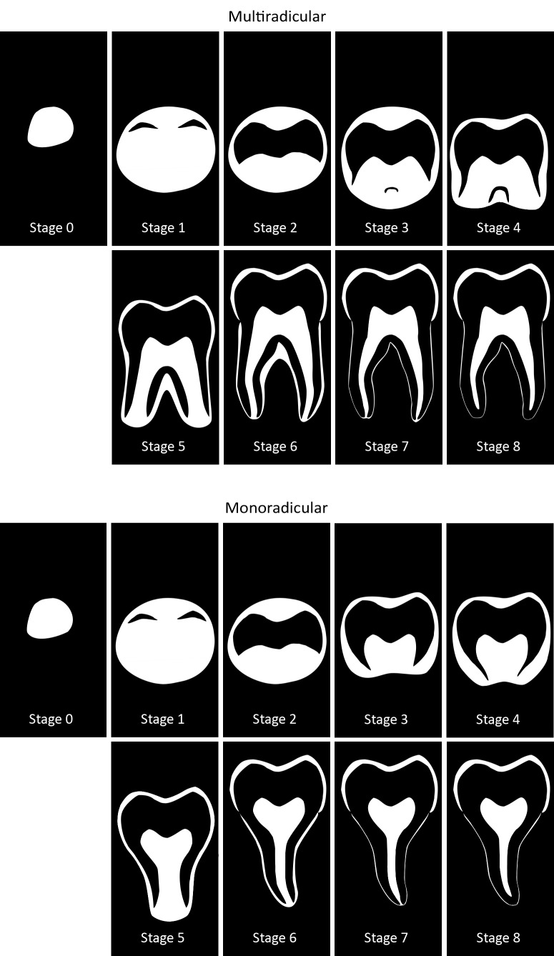
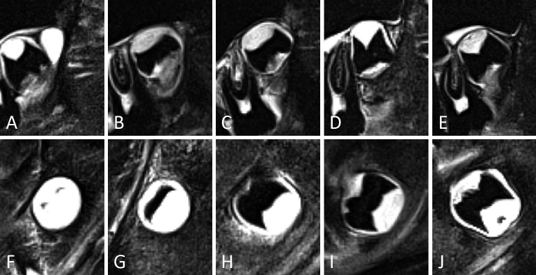
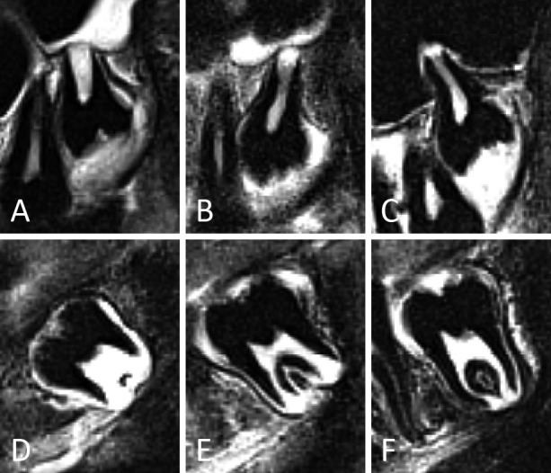
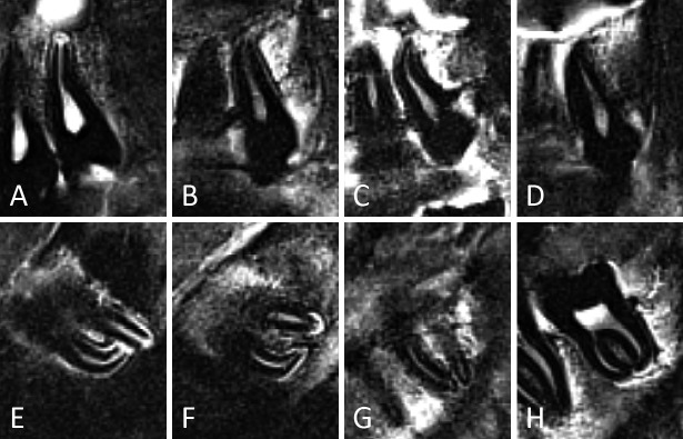
Background: The development of third molars can be evaluated with medical imaging to estimate age in subadults. The appearance of third molars on magnetic resonance imaging (MRI) differs greatly from that on radiographs. Therefore a specific staging technique is necessary to classify third molar development on MRI and to apply it for age estimation.
Aim: To develop a specific staging technique to register third molar development on MRI and to evaluate its performance for age estimation in subadults.
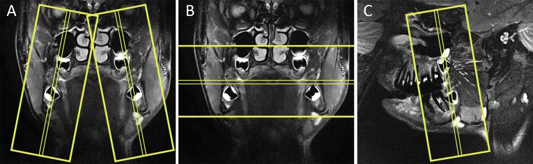
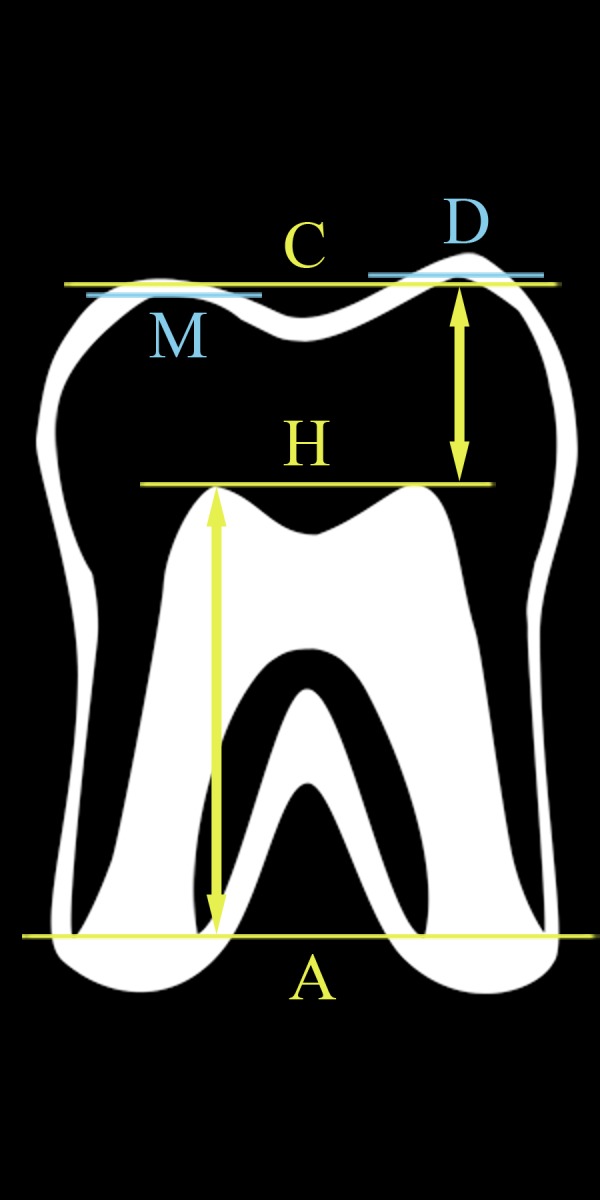
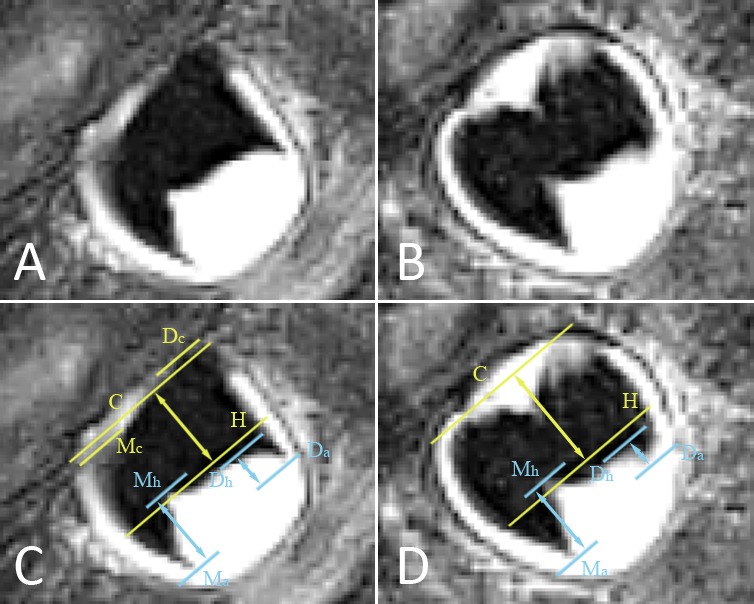
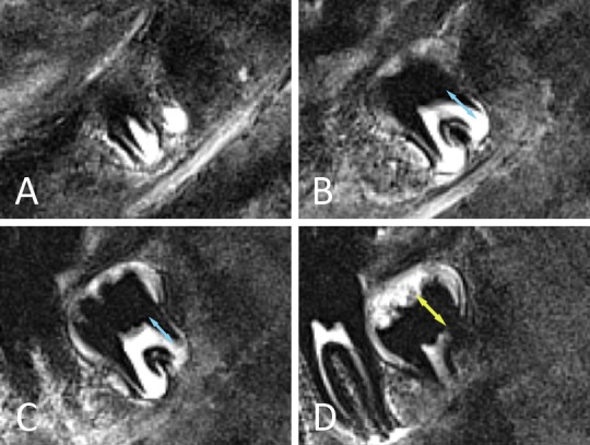
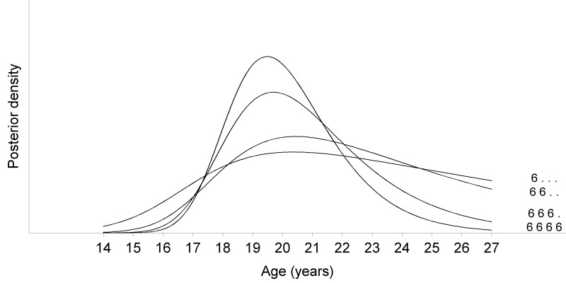
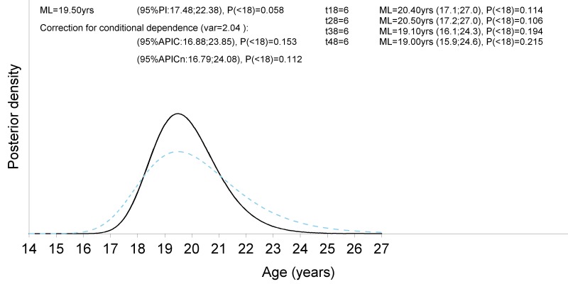
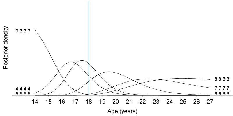
Materials and methods: Using 3T MRI in three planes, all third molars were evaluated in 309 healthy Caucasian participants from 14 to 26 years old. According to the appearance of the developing third molars on MRI, descriptive criteria and schematic representations were established to define a specific staging technique. Two observers, with different levels of experience, staged all third molars independently with the developed technique. Intra- and inter-observer agreement were calculated. The data were imported in a Bayesian model for age estimation as described by Fieuws et al. (2016). This approach adequately handles correlation between age indicators and missing age indicators. It was used to calculate a point estimate and a prediction interval of the estimated age. Observed age minus predicted age was calculated, reflecting the error of the estimate.
Results: One-hundred and sixty-six third molars were agenetic. Five percent (51/1096) of upper third molars and 7% (70/1044) of lower third molars were not assessable. Kappa for inter-observer agreement ranged from 0.76 to 0.80. For intra-observer agreement kappa ranged from 0.80 to 0.89. However, two stage differences between observers or between staging sessions occurred in up to 2.2% (20/899) of assessments, probably due to a learning effect. Using the Bayesian model for age estimation, a mean absolute error of 2.0 years in females and 1.7 years in males was obtained. Root mean squared error equalled 2.38 years and 2.06 years respectively. The performance to discern minors from adults was better for males than for females, with specificities of 96% and 73% respectively.
Conclusion: Age estimations based on the proposed staging method for third molars on MRI showed comparable reproducibility and performance as the established methods based on radiographs.
Conflict of interest statement
Conflicts of Interest: None declared.
Figures

Similar articles
-
Forensic age estimation based on magnetic resonance imaging of third molars: converting 2D staging into 3D staging.Ann Hum Biol. 2017 Mar;44(2):121-129. doi: 10.1080/03014460.2016.1223884. Epub 2016 Sep 20. Ann Hum Biol. 2017. PMID: 27650301
-
An automated technique to stage lower third molar development on panoramic radiographs for age estimation: a pilot study.J Forensic Odontostomatol. 2017 Dec 1;35(2):42-54. J Forensic Odontostomatol. 2017. PMID: 29384736 Free PMC article.
-
Applicability of Willems model for dental age estimations in Brazilian children.Forensic Sci Int. 2013 Sep 10;231(1-3):401.e1-4. doi: 10.1016/j.forsciint.2013.05.030. Epub 2013 Jun 24. Forensic Sci Int. 2013. PMID: 23806342
-
Third-molar development in relation to chronologic age in Turkish children and young adults.Angle Orthod. 2007 Nov;77(6):1040-5. doi: 10.2319/101906-430.1. Angle Orthod. 2007. PMID: 18004924 Review.
-
Dental age estimation utilizing third molar development: A review of principles, methods, and population studies used in the United States.Forensic Sci Int. 2010 Sep 10;201(1-3):79-83. doi: 10.1016/j.forsciint.2010.04.042. Epub 2010 May 20. Forensic Sci Int. 2010. PMID: 20493649 Review.
Cited by
-
CT and MR imaging used in age estimation: a systematic review.J Forensic Odontostomatol. 2018 May 30;36(1):14-25. J Forensic Odontostomatol. 2018. PMID: 29864026 Free PMC article.
-
Magnetic Resonance Images for the Prediction of Chronological Age Using Maxillary Third Molar Teeth: An Observational Study.Clin Cosmet Investig Dent. 2024 Oct 15;16:405-411. doi: 10.2147/CCIDE.S484107. eCollection 2024. Clin Cosmet Investig Dent. 2024. PMID: 39429438 Free PMC article.
-
Inter-observer agreement in the radiographic interpretation of Demirjian's developmental stages in the mandibular second and third molars - A comparative study.J Oral Maxillofac Pathol. 2021 Sep-Dec;25(3):554-555. doi: 10.4103/jomfp.jomfp_85_21. Epub 2022 Jan 11. J Oral Maxillofac Pathol. 2021. PMID: 35281143 Free PMC article.
-
Age estimation based on 3D post-mortem computed tomography images of mandible and femur using convolutional neural networks.PLoS One. 2021 May 12;16(5):e0251388. doi: 10.1371/journal.pone.0251388. eCollection 2021. PLoS One. 2021. PMID: 33979376 Free PMC article.
-
Systematic review: oral and maxillofacial radiology as fundamental methods of virtual autopsy.Forensic Sci Res. 2023 Nov 10;8(3):185-197. doi: 10.1093/fsr/owad028. eCollection 2023 Sep. Forensic Sci Res. 2023. PMID: 39633891 Free PMC article. Review.
References
MeSH terms
LinkOut - more resources
Full Text Sources
Medical