Spontaneous cerebrospinal fluid rhinorrhea: A case report and analysis
- PMID: 29384861
- PMCID: PMC5805433
- DOI: 10.1097/MD.0000000000009758
Spontaneous cerebrospinal fluid rhinorrhea: A case report and analysis
Erratum in
-
Spontaneous cerebrospinal fluid rhinorrhea: A case report and analysis: Erratum.Medicine (Baltimore). 2018 Feb;97(7):e9954. doi: 10.1097/MD.0000000000009954. Medicine (Baltimore). 2018. PMID: 29443787 Free PMC article. No abstract available.
Abstract
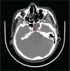
Introduction: Spontaneous cerebrospinal fluid leakage is usually caused by developmental abnormalities and is rare, accounting for approximately 5% of the cases of cerebrospinal fluid (CSF) leakage. To the best of our knowledge, clival dysplasia-caused CSF rhinorrhea has never been reported in the neurosurgical field.
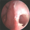
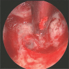
Conclusion: Spontaneous cerebrospinal fluid rhinorrhea is often treated by surgery, and a transsphenoidal approach repair is the main surgical method used, offering the advantages of less trauma, fewer complications, rapid postoperative recovery, and low recurrence rate.
Conflict of interest statement
The authors have no conflicts of interest to disclose.
Figures


Similar articles
-
Anterior Clival Defect with Primary CSF Rhinorrhea: A Case Report.Neurol India. 2022 Jan-Feb;70(1):352-354. doi: 10.4103/0028-3886.338711. Neurol India. 2022. PMID: 35263912
-
Endoscopic Endonasal Repair of Cerebrospinal Fluid Leakage Caused by a Rare Traumatic Clival Fracture.Neurol Med Chir (Tokyo). 2016;56(2):81-4. doi: 10.2176/nmc.cr.2015-0152. Epub 2016 Jan 22. Neurol Med Chir (Tokyo). 2016. PMID: 26804187 Free PMC article.
-
Recurrent Spontaneous Cerebrospinal Fluid Rhinorrhea With Meningitis due to a Clival Defect and Literature Review.J Craniofac Surg. 2025 Sep 1;36(6):2049-2051. doi: 10.1097/SCS.0000000000011681. Epub 2025 Jul 25. J Craniofac Surg. 2025. PMID: 40737103 Review.
-
Spontaneous CSF rhinorrhea as a presenting symptom of a clival chordoma.Neurocirugia (Engl Ed). 2021 Jan-Feb;32(1):41-43. doi: 10.1016/j.neucir.2019.12.002. Epub 2020 Jan 27. Neurocirugia (Engl Ed). 2021. PMID: 32001132 English, Spanish.
-
Treating Cerebrospinal Fluid Rhinorrhea without Dura Repair: A Case Report of Posterior Fossa Choroid Plexus Papilloma and Review of the Literature.World Neurosurg. 2017 Dec;108:990.e1-990.e9. doi: 10.1016/j.wneu.2017.08.121. Epub 2017 Sep 1. World Neurosurg. 2017. PMID: 28866068 Review.
Cited by
-
Spontaneous cerebrospinal fluid rhinorrhea: A case report and analysis: Erratum.Medicine (Baltimore). 2018 Mar;97(12):e0274. doi: 10.1097/MD.0000000000010274. Medicine (Baltimore). 2018. PMID: 29561458 Free PMC article. No abstract available.
-
Cerebrospinal fluid rhinorrhea in a bilateral frontal decompressive craniectomy patient caused by strenuous activity: A case report.Medicine (Baltimore). 2018 Nov;97(47):e13189. doi: 10.1097/MD.0000000000013189. Medicine (Baltimore). 2018. PMID: 30461618 Free PMC article.
-
Spontaneous cerebrospinal fluid rhinorrhea: A case report and analysis: Erratum.Medicine (Baltimore). 2018 Feb;97(7):e9954. doi: 10.1097/MD.0000000000009954. Medicine (Baltimore). 2018. PMID: 29443787 Free PMC article. No abstract available.
-
Spontaneous cerebrospinal fluid rhinorrhoea: a case report and literature review.J Med Case Rep. 2024 Nov 11;18(1):533. doi: 10.1186/s13256-024-04882-9. J Med Case Rep. 2024. PMID: 39523355 Free PMC article. Review.
-
A Rare Case of Cerebrospinal Fluid Rhinorrhea from Canal of Stenberg.Indian J Otolaryngol Head Neck Surg. 2023 Apr;75(Suppl 1):764-767. doi: 10.1007/s12070-022-03347-z. Epub 2022 Dec 11. Indian J Otolaryngol Head Neck Surg. 2023. PMID: 37206705 Free PMC article.
References
-
- Fraioli MF, Umana GE, Fiorucci G, et al. Ethmoidal encephalocele associated with cerebrospinal fluid fistula: indications and results of mini-invasive transnasal approach. J Craniofac Surg 2014;25:551–3. - PubMed
-
- Reddy M, Baugnon K. Imaging of Cerebrospinal Fluid Rhinorrhea and Otorrhea. Radiol Clin North Am 2017;55:167–87. - PubMed
-
- Tumturk A, Ulutabanca H, Gokoglu A, et al. The results of the surgical treatment of spontaneous rhinorrhea via craniotomy and the contribution of CT cisternography to the detection of exact leakage side of CSF. Turk Neurosurg 2016;26:699–703. - PubMed
-
- Connor SE. Imaging of skull-base cephalocoeles and cerebrospinal fluid leaks. Clin Radiol 2010;65:832–41. - PubMed
Publication types
MeSH terms
LinkOut - more resources
Full Text Sources
Other Literature Sources

