

Polymeric Micelle of A₃B-Type Lactosome as a Vehicle for Targeting Meningeal Dissemination
- PMID: 29385027
- PMCID: PMC5853711
- DOI: 10.3390/nano8020079
Polymeric Micelle of A₃B-Type Lactosome as a Vehicle for Targeting Meningeal Dissemination
Abstract
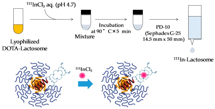
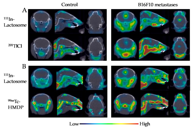
Polymeric micelle of the A₃B-type lactosome comprising (poly(sarcosine))₃-b-poly(l-lactic acid) was labeled with 111In. The 111In-labeled A₃B-type lactosome was administered to the model mice bearing meningeal dissemination and bone metastasis at mandible. With single-photon emission computed tomography (SPECT) imaging, the meningeal dissemination was identified successfully by 111In-labeled A₃B-type lactosome, which was superior to 201TlCl in regard of the imaging contrast. The 111In-labeled A₃B-type lactosome was also potential in imaging selectively of bone metastasis at mandible, whilst a nonspecific imaging of the whole bone was obtained by the SPECT imaging using 99mTc-HMDP. The polymeric micelle of the A₃B-type lactosome was therefore found to be effective as a vehicle of 111In to be targeted to meningeal dissemination and bone metastasis.
Keywords: SPECT; bone metastasis; meningeal dissemination; molecular imaging; polymeric micelle.
Conflict of interest statement
The authors declare no conflict of interest.
Figures


Similar articles
-
A Novel 89Zr-labeled DDS Device Utilizing Human IgG Variant (scFv): "Lactosome" Nanoparticle-Based Theranostics for PET Imaging and Targeted Therapy.Life (Basel). 2021 Feb 18;11(2):158. doi: 10.3390/life11020158. Life (Basel). 2021. PMID: 33670777 Free PMC article. Review.
-
Radiosynthesis and initial evaluation of (18)F labeled nanocarrier composed of poly(L-lactic acid)-block-poly(sarcosine) amphiphilic polydepsipeptide.Nucl Med Biol. 2013 Apr;40(3):387-94. doi: 10.1016/j.nucmedbio.2012.12.008. Epub 2013 Jan 22. Nucl Med Biol. 2013. PMID: 23347829
-
18F-Labeled poly(L-lactic acid)-block-poly(sarcosine) lactosome, a polymer micelle.2013 Apr 5 [updated 2013 May 2]. In: Molecular Imaging and Contrast Agent Database (MICAD) [Internet]. Bethesda (MD): National Center for Biotechnology Information (US); 2004–2013. 2013 Apr 5 [updated 2013 May 2]. In: Molecular Imaging and Contrast Agent Database (MICAD) [Internet]. Bethesda (MD): National Center for Biotechnology Information (US); 2004–2013. PMID: 23638489 Free Books & Documents. Review.
-
Factors influencing in vivo disposition of polymeric micelles on multiple administrations.ACS Med Chem Lett. 2014 Jun 18;5(8):873-7. doi: 10.1021/ml500112u. eCollection 2014 Aug 14. ACS Med Chem Lett. 2014. PMID: 25147606 Free PMC article.
-
Activation of B1a cells in peritoneal cavity by T cell-independent antigen expressed on polymeric micelle.J Pharm Sci. 2015 May;104(5):1839-47. doi: 10.1002/jps.24397. Epub 2015 Feb 26. J Pharm Sci. 2015. PMID: 25720375
Cited by
-
A Novel 89Zr-labeled DDS Device Utilizing Human IgG Variant (scFv): "Lactosome" Nanoparticle-Based Theranostics for PET Imaging and Targeted Therapy.Life (Basel). 2021 Feb 18;11(2):158. doi: 10.3390/life11020158. Life (Basel). 2021. PMID: 33670777 Free PMC article. Review.
-
Biodistribution of 68/67Ga-Radiolabeled Sphingolipid Nanoemulsions by PET and SPECT Imaging.Int J Nanomedicine. 2021 Aug 26;16:5923-5935. doi: 10.2147/IJN.S316767. eCollection 2021. Int J Nanomedicine. 2021. PMID: 34475757 Free PMC article.
-
Recent Status of Nanomaterial Fabrication and Their Potential Applications in Neurological Disease Management.Nanoscale Res Lett. 2018 Aug 10;13(1):231. doi: 10.1186/s11671-018-2638-7. Nanoscale Res Lett. 2018. PMID: 30097809 Free PMC article. Review.
References
-
- Balch C.M., Soong S.J., Gershenwald J.E., Thompson J.F., Reintgen D.S., Cascinelli N., Urist M., McMasters K.M., Ross M.I., Kirkwood J.M., et al. Prognostic factors analysis of 17,600 melanoma patients: Validation of the American joint committee on cancer melanoma staging system. J. Clin. Oncol. 2001;19:3622–3634. doi: 10.1200/JCO.2001.19.16.3622. - DOI - PubMed
LinkOut - more resources
Full Text Sources
Other Literature Sources
