A Pathological Study of Acute Pulmonary Toxicity Induced by Inhaled Kanto Loam Powder
- PMID: 29385040
- PMCID: PMC5855638
- DOI: 10.3390/ijms19020416
A Pathological Study of Acute Pulmonary Toxicity Induced by Inhaled Kanto Loam Powder
Abstract
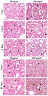
The frequency and volume of Asian sand dust (ASD) (Kosa) are increasing in Japan, and it has been reported that ASD may cause adverse respiratory effects. The pulmonary toxicity of ASD has been previously analyzed in mice exposed to ASD particles by intratracheal instillation. To study the pulmonary toxicity induced by inhalation of ASD, ICR mice were exposed by inhalation to 50 or 200 mg/m³ Kanto loam powder, which resembles ASD in elemental composition and particle size, for 6 h a day over 1, 3, 6, 9, or 15 consecutive days. Histological examination revealed that Kanto loam powder induced acute inflammation in the whole lung at all the time points examined. The lesions were characterized by infiltration of neutrophils and macrophages. The intensity of the inflammatory changes in the lung and number of neutrophils in both histological lesions and bronchoalveolar lavage fluid (BALF) appeared to increase over time. Immunohistochemical staining showed interleukin (IL)-6- and tumor necrosis factor (TNF)-α-positive macrophages and a decrease in laminin positivity in the inflammatory lesions of the lung tissues. Electron microscopy revealed vacuolar degeneration in the alveolar epithelial cells close to the Kanto loam particles. The nitric oxide level in the BALF increased over time. These results suggest that inhaled Kanto loam powder may induce diffuse and acute pulmonary inflammation, which is associated with increased expression of inflammatory cytokines and oxidative stress.
Keywords: Asian sand dust; Kanto loam powder; inhalation; pulmonary toxicity.
Conflict of interest statement
The authors declare no conflict of interest. The founding sponsors had no role in the design of the study; in the collection, analyses, or interpretation of data; in the writing of the manuscript; or in the decision to publish the results.
Figures


Similar articles
-
Pathological study of chronic pulmonary toxicity induced by intratracheally instilled Asian sand dust (kosa).Toxicol Pathol. 2013 Jan;41(1):48-62. doi: 10.1177/0192623312452490. Epub 2012 Jun 28. Toxicol Pathol. 2013. PMID: 22744225
-
Pathological study of acute pulmonary toxicity induced by intratracheally instilled Asian sand dust (kosa).Toxicol Pathol. 2010 Dec;38(7):1099-110. doi: 10.1177/0192623310385143. Epub 2010 Sep 30. Toxicol Pathol. 2010. PMID: 20884819
-
Pathological study of pulmonary toxicity induced by intratracheally instilled Asian sand dust (Kosa): effects of lowered serum zinc level on the toxicity.Folia Histochem Cytobiol. 2018;56(1):38-48. doi: 10.5603/FHC.a2018.0006. Epub 2018 Mar 26. Folia Histochem Cytobiol. 2018. PMID: 29577227
-
Asian sand dust enhances murine lung inflammation caused by Klebsiella pneumoniae.Toxicol Appl Pharmacol. 2012 Jan 15;258(2):237-47. doi: 10.1016/j.taap.2011.11.003. Epub 2011 Nov 19. Toxicol Appl Pharmacol. 2012. PMID: 22118940
-
Is pulmonary inflammation a valid predictor of particle induced lung pathology? The case of amorphous and crystalline silicas.Toxicol Lett. 2024 Aug;399 Suppl 1:18-30. doi: 10.1016/j.toxlet.2023.07.012. Epub 2023 Jul 14. Toxicol Lett. 2024. PMID: 37454774 Review.
Cited by
-
TXNIP regulates pulmonary inflammation induced by Asian sand dust.Redox Biol. 2024 Dec;78:103421. doi: 10.1016/j.redox.2024.103421. Epub 2024 Nov 6. Redox Biol. 2024. PMID: 39520910 Free PMC article.
References
-
- Kurosaki Y., Shinoda M., Mikami M. What caused a recent increase in dust outbreaks over East Asia? Geophys. Res. Lett. 2011;38:L11702. doi: 10.1029/2011GL047494. - DOI
-
- Lin C.-Y., Wang Z., Chen W.-N., Chang S.-Y., Chou C.C.K., Sugimoto N., Zhao X. Long-range transport of Asian dust and air pollutants to Taiwan: Observed evidence and model simulation. Atmos. Chem. Phys. 2007;7:423–434. doi: 10.5194/acp-7-423-2007. - DOI
-
- Lee H., Kim H., Honda Y., Lim Y.H., Yi S. Effect of Asian dust storms on daily mortality in seven metropolitan cities of Korea. Atmos. Environ. 2013;79:510–517. doi: 10.1016/j.atmosenv.2013.06.046. - DOI
MeSH terms
Substances
LinkOut - more resources
Full Text Sources
Other Literature Sources
Medical

