. 2018 Jan 31;19(2):421.
doi: 10.3390/ijms19020421.

Implementation of Glycan Remodeling to Plant-Made Therapeutic Antibodies

Affiliations

Implementation of Glycan Remodeling to Plant-Made Therapeutic Antibodies

Lindsay D Bennett et al. Int J Mol Sci. .

Abstract

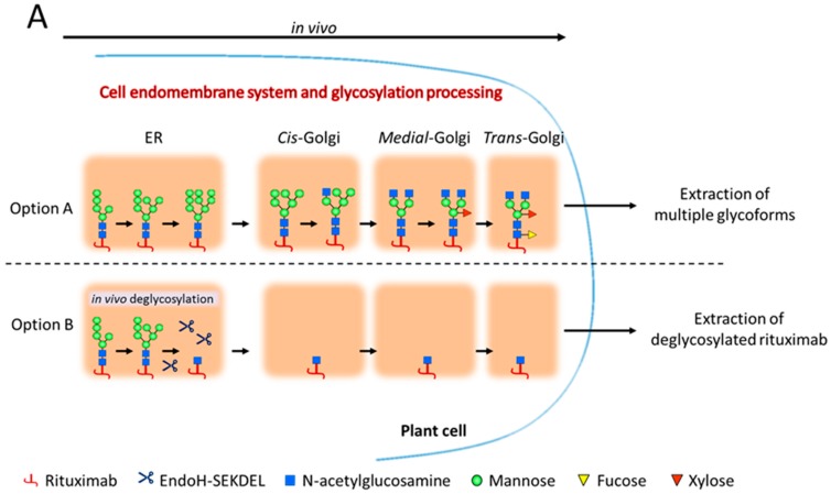
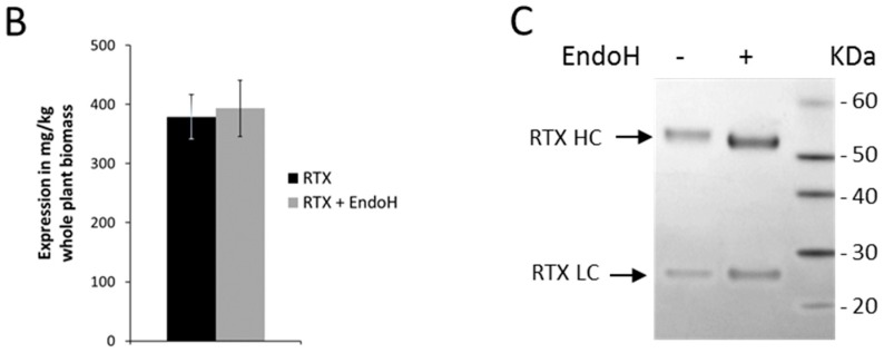
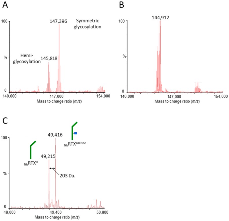
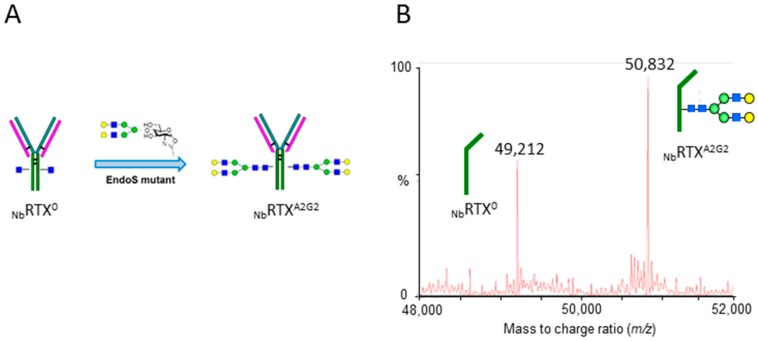
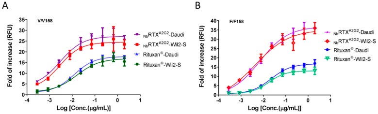
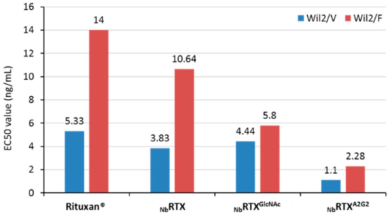
N-glycosylation profoundly affects the biological stability and function of therapeutic proteins, which explains the recent interest in glycoengineering technologies as methods to develop biobetter therapeutics. In current manufacturing processes, N-glycosylation is host-specific and remains difficult to control in a production environment that changes with scale and production batches leading to glycosylation heterogeneity and inconsistency. On the other hand, in vitro chemoenzymatic glycan remodeling has been successful in producing homogeneous pre-defined protein glycoforms, but needs to be combined with a cost-effective and scalable production method. An efficient chemoenzymatic glycan remodeling technology using a plant expression system that combines in vivo deglycosylation with an in vitro chemoenzymatic glycosylation is described. Using the monoclonal antibody rituximab as a model therapeutic protein, a uniform Gal2GlcNAc2Man3GlcNAc2 (A2G2) glycoform without α-1,6-fucose, plant-specific α-1,3-fucose or β-1,2-xylose residues was produced. When compared with the innovator product Rituxan®, the plant-made remodeled afucosylated antibody showed similar binding affinity to the CD20 antigen but significantly enhanced cell cytotoxicity in vitro. Using a scalable plant expression system and reducing the in vitro deglycosylation burden creates the potential to eliminate glycan heterogeneity and provide affordable customization of therapeutics' glycosylation for maximal and targeted biological activity. This feature can reduce cost and provide an affordable platform to manufacture biobetter antibodies.

Keywords: N-glycosylation; Nicotiana benthamiana; glycan remodeling; recombinant glycoproteins; therapeutic proteins.


Conflict of interest statement

The authors declare no conflict of interest.

Figures

Figure 1
Figure 1
(A) Schematic representation of in vivo N-glycan processing of recombinant proteins in standard plant expression systems (Option A) and when co-expressed with EndoH (Option B). The Option B pathway illustrates the in vivo deglycosylation strategy employed for rituximab custom glycosylation where high-mannose glycans attached to the protein of interest are cleaved off by EndoH, leaving a deglycosylated substrate for in vitro transglycosylation; (B) plots of rituximab expression levels in mg/kg of plant biomass. Protein was harvested from plants expressing rituximab alone (black bar) or coexpressing rituximab with EndoH (grey bar) (Values are means ± SEM, n = 4); (C) SDS-PAGE showing rituximab under reduced conditions, with or without EndoH coexpression. The light chain (RTX LC) species for both samples migrated similarly, while the heavy chain (RTX HC) from plants coexpressing EndoH underwent an increased mobility relative to the rituximab alone, indicating a decrease in molecular weight.
Figure 1
Figure 1
(A) Schematic representation of in vivo N-glycan processing of recombinant proteins in standard plant expression systems (Option A) and when co-expressed with EndoH (Option B). The Option B pathway illustrates the in vivo deglycosylation strategy employed for rituximab custom glycosylation where high-mannose glycans attached to the protein of interest are cleaved off by EndoH, leaving a deglycosylated substrate for in vitro transglycosylation; (B) plots of rituximab expression levels in mg/kg of plant biomass. Protein was harvested from plants expressing rituximab alone (black bar) or coexpressing rituximab with EndoH (grey bar) (Values are means ± SEM, n = 4); (C) SDS-PAGE showing rituximab under reduced conditions, with or without EndoH coexpression. The light chain (RTX LC) species for both samples migrated similarly, while the heavy chain (RTX HC) from plants coexpressing EndoH underwent an increased mobility relative to the rituximab alone, indicating a decrease in molecular weight.
Figure 2
Figure 2
Nano liquid chromatography-electrospray ionization-quadrupole time of flight-mass spectrometry (NanoLC-QTOF-MS) analysis of plant-made rituximab. (A) Deconvoluted electrospray ionization (ESI)-mass spectrum of purified NbRTX analyzed under non-reducing conditions showing fully glycosylated and hemiglycosylated rituximab; (B) Deconvoluted ESI-mass spectrum of purified NbRTXGlcNAc analyzed under non-reducing conditions showing a decrease in molecular weight; (C) Deconvoluted ESI-mass spectrum of in vivo deglycosylated plant-made rituximab analyzed under reducing conditions. Note the mass shift between non-glycosylated rituximab heavy chain NbRTX0 (calculated m/z 49,217) and deglycosylated rituximab heavy chain NbRTXGlcNAc (calculated m/z 49,420). As comparison, the NanoLC-QTOF-MS analysis of Rituxan® is provided in Figure S2.
Figure 3
Figure 3
Chemoenzymatic transglycosylation of plant-made rituximab. (A) Schematic representation of the chemoenzymatic transglycosylation reaction; (B) NanoLC-QTOF-MS analysis of reglycosylated plant-made rituximab. Deconvoluted ESI-mass spectrum of reglycosylated plant-made rituximab with the A2G2 glycan (NbRTXA2G2 heavy chain, calculated m/z 50,840) analyzed under reducing conditions.
Figure 4
Figure 4
Binding of Rituxan® (A,B) and NbRTXA2G2 (C,D) to Wil2-S and Daudi cells analyzed by flow cytometry. Rituxan® and NbRTXA2G2 were used at a concentration of 10 nM. All rituximab samples were measured in triplicates (red, green and blue lines). FITC-labeled mouse IgG2a (black line) and unstained cells (grey line) were used as controls. The X-axis represents the fluorescent signals of FITC whereas the Y-axis presents % of cell count.
Figure 4
Figure 4
Binding of Rituxan® (A,B) and NbRTXA2G2 (C,D) to Wil2-S and Daudi cells analyzed by flow cytometry. Rituxan® and NbRTXA2G2 were used at a concentration of 10 nM. All rituximab samples were measured in triplicates (red, green and blue lines). FITC-labeled mouse IgG2a (black line) and unstained cells (grey line) were used as controls. The X-axis represents the fluorescent signals of FITC whereas the Y-axis presents % of cell count.
Figure 5
Figure 5
Antibody-dependent-cell-mediated cytotoxicity (ADCC) activity of Rituxan® and NbRTXA2G2 with (A) V/V 158 FcγRIIIa (high affinity) and (B) F/F 158 FcγRIIIa (low affinity) variant effector cells (engineered Jurkat cells with FcγRIIIa receptor). All experiments were carried out using human B lymphoma WIL2-S cells and Daudi cells as target cells. The effector cell: target cell ratio was 10:1. Data are expressed as fold of ADCC increase. Values represent mean ± S.D. for triplicate analyses.
Figure 6
Figure 6
Comparison of EC50 values of Rituxan®, plant-made rituximab (NbRTX), deglycosylated plant-made rituximab (NbRTXGlcNAc), and reglycosylated plant-made rituximab (NbRTXA2G2) determined by the ADCC Reporter Bioassay using Wil2 V/V158 (Wil2/V) and F/F158 (Wil2/F) variant cells. The calculated EC50 values are shown above the respective histogram bars.

References

    1. Walsh G., Jefferis R. Post-translational modifications in the context of therapeutic proteins. Nat. Biotechnol. 2006;24:1241–1252. doi: 10.1038/nbt1252. - DOI - PubMed
    1. Costa A.R., Rodrigues M.E., Henriques M., Oliveira R., Azeredo J. Glycosylation: Impact, control and improvement during therapeutic protein production. Crit. Rev. Biotechnol. 2013;34:281–299. doi: 10.3109/07388551.2013.793649. - DOI - PubMed
    1. Hossler P., Khattak S.F., Li Z.J. Optimal and consistent protein glycosylation in mammalian cell culture. Glycobiology. 2009;19:936–949. doi: 10.1093/glycob/cwp079. - DOI - PubMed
    1. Li H., D’Anjou M. Pharmacological significance of glycosylation in therapeutic proteins. Curr. Opin. Biotechnol. 2009;20:678–684. doi: 10.1016/j.copbio.2009.10.009. - DOI - PubMed
    1. Jefferis R. Glyco-Engineering of Human IgG-Fc to Modulate Biologic Activities. Curr. Pharm. Biotechnol. 2016;17:1333–1347. doi: 10.2174/1389201017666161029225929. - DOI - PubMed