Review
. 2018 Jan 31;23(2):301.
doi: 10.3390/molecules23020301.

Pineal Calcification, Melatonin Production, Aging, Associated Health Consequences and Rejuvenation of the Pineal Gland

Affiliations
Review

Pineal Calcification, Melatonin Production, Aging, Associated Health Consequences and Rejuvenation of the Pineal Gland

Dun Xian Tan et al. Molecules. .

Abstract

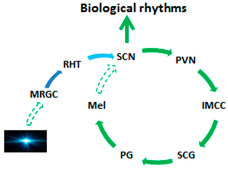
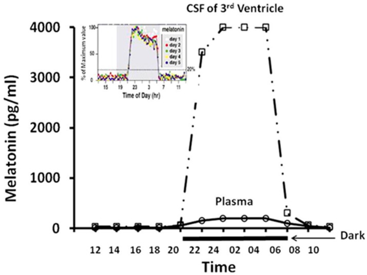
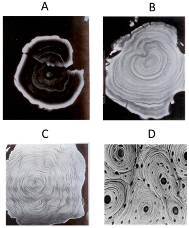
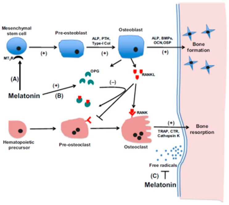
The pineal gland is a unique organ that synthesizes melatonin as the signaling molecule of natural photoperiodic environment and as a potent neuronal protective antioxidant. An intact and functional pineal gland is necessary for preserving optimal human health. Unfortunately, this gland has the highest calcification rate among all organs and tissues of the human body. Pineal calcification jeopardizes melatonin's synthetic capacity and is associated with a variety of neuronal diseases. In the current review, we summarized the potential mechanisms of how this process may occur under pathological conditions or during aging. We hypothesized that pineal calcification is an active process and resembles in some respects of bone formation. The mesenchymal stem cells and melatonin participate in this process. Finally, we suggest that preservation of pineal health can be achieved by retarding its premature calcification or even rejuvenating the calcified gland.

Keywords: aging; calcification; melatonin; neurodegenerative diseases; pineal gland; rejuvenation.


Conflict of interest statement

The authors declare no conflict of interest.

Figures

Figure 1
Figure 1
Illustration of the SCN-melatonin loop. Solid arrows indicate the neuronal connections and the direction of neuronal projections. Dash arrows indicate the input signals. SCN is the master clock which determines the biological rhythms as well as the melatonin circadian rhythm. Its intrinsic circadian interval is longer than 24 h. The natural photoperiod (photos) serves as an input signal to entrain melatonin circadian rhythm to 24 h; in turn, melatonin functions as a signal of photoperiod to re-entrain the biological rhythm of SCN to 24 h. MRGC: melanopsin-containing retinal ganglion cells; RHT: retino-hypothalamic tract; SCN: suprachasmatic nucleus; PVN: paraventricular nucleus; IMCC: Intermediolateral cell column; SCG: sympathetic cervical ganglion; PG: pineal gland. Mel: melatonin.
Figure 2
Figure 2
The different levels and shapes of melatonin circadian rhythms in the CSF of the third ventricle and in the peripheral blood. The nightly melatonin levels in CSF of the third ventricle are more robust than those in the peripheral plasma and also exhibit sharp rises and falls (square wave). The pattern of the melatonin circadian rhythm in CSF of the third ventricle is similar to that of the pineal gland rather than in the plasma (see the insert part which illustrates the melatonin synthetic pattern in pineal gland of rat). The data was obtained from the long term (5 days) pineal gland dialysis in a free running rat. Extrapineal-generated melatonin and the diet-derived melatonin may increase peripheral plasma melatonin levels; however, they do not mimic the pattern and reach the high level of the melatonin circadian rhythm in the CSF of the third ventricle to impact the function of bio-clock. From Tan et al. [63].
Figure 3
Figure 3
The laminated pineal gland calcification at different ages and their similarity to the osteons of compact bone. (A) Pineal calcification in 14 year old subject; (B) 47 year old; (C) 62 year old; (D) osteons; (AC) were modified from Hermann et al. [239].
Figure 4
Figure 4
The proposed mechanisms underlying melatonin’s actions on bone formation. (A) melatonin induces MSCs differentiation into osteoblasts via MT2; (B) it promotes osteoprotegerin (OPG) expression in preosteoblasts which would inactive RANKL, leading to a suppression of osteoclastogenesis; and (C) through melatonin’s free-radical scavenging and antioxidant properties, protecting against radical induced loss of osteoblasts and osteoclasts. PTH (parathyroid hormone); Type I col (type I collagen); OSP (osteopontin); BMP-2 (bone morphogenetic protein 2); ALP (alkaline phosphatase); OCN (osteocalcin); TRAP (tartrate-resistant acid phosphatase); RANKL (receptor activator of NFĸB ligand); OPG (osteoprotegerin). From Maria and Witt-Enderby [262].
Figure 5
Figure 5
Histology and ultrastructure of cells located in a concretion of the turkey pineal gland. (A) mature concretions identified with Mallory’s stain. Two types of cells are present in the concretion (semi-thin section stained with toluidine blue); (B) polygonal cells; (C) elongated cells; (D) The presence of calcium in the concretions was demonstrated with Alizarin red S; note the characteristic appearance of cells located in the concretion; (E) ultrastructure of cells located in the calcified area (fixation with the PPA method). 1: the osteocyte-like cell surrounded by mineralized collagen fibrils in the central part of the calcification area. Note the “halo” around the cell and large pyroantimonate precipitate located mainly outside the cell membrane, 2: the junction of processes of osteocyte-like cells, 3: a cell showing a fibrocyte-like appearance in the peripheral part of the calcification area. Note the extra-cellular matrix containing collagen and calcium deposits, 4: numerous calcium precipitates in the intercellular spaces in the peripheral part of the calcification area, 5: The cell process with scattered deposits in the middle part of the concretion. Note the adjacent extra-cellular matrix rich in collagen and pyroantimonate precipitates. Modified from Przybylska-Gornowicz et al. [159].

References

    1. Golan J., Torres K., Staśkiewicz G.J., Opielak G., Maciejewski R. Morphometric parameters of the human pineal gland in relation to age, body weight and height. Folia Morphol. 2002;61:111–113. - PubMed
    1. Tan D.X., Manchester L.C., Fuentes-Broto L., Paredes S.D., Reiter R.J. Significance and application of melatonin in the regulation of brown adipose tissue metabolism: Relation to human obesity. Obes. Rev. 2011;12:167–188. doi: 10.1111/j.1467-789X.2010.00756.x. - DOI - PubMed
    1. Hintermann E., Jenö P., Meyer U.A. Isolation and characterization of an arylalkylamine N-acetyltransferase from Drosophila melanogaster. FEBS Lett. 1995;375:148–150. doi: 10.1016/0014-5793(95)01198-N. - DOI - PubMed
    1. Itoh M.T., Hattori A., Sumi Y., Suzuki T. Day-night changes in melatonin levels in different organs of the cricket (Gryllus bimaculatus) J. Pineal Res. 1995;18:165–169. doi: 10.1111/j.1600-079X.1995.tb00156.x. - DOI - PubMed
    1. Vivien-Roels B., Pevet P., Beck O., Fevre-Montange M. Identification of melatonin in the compound eyes of an insect, the locust (Locusta migratoria), by radioimmunoassay and gas chromatography-mass spectrometry. Neurosci. Lett. 1984;49:153–157. doi: 10.1016/0304-3940(84)90152-6. - DOI - PubMed