. 2018 May;33(5):555-568.
doi: 10.1002/tox.22541. Epub 2018 Jan 31.

Toxicity implications for early life stage Japanese medaka (Oryzias latipes) exposed to oxyfluorfen

Affiliations

Toxicity implications for early life stage Japanese medaka (Oryzias latipes) exposed to oxyfluorfen

Doris K Powe et al. Environ Toxicol. 2018 May.

Abstract

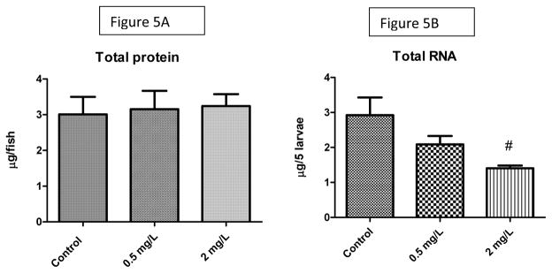
We investigated the potential toxic effects of Oxyfluorfen (OXY), an herbicide used in agriculture, on the embryo-larval development of Japanese medaka fish (Oryzias latipes). Embryos (1-day postfertilization) and larvae (2-day posthatch) were exposed to OXY (0.5-8 mg/L) for 96 h and evaluated for mortality and hatching on embryos, and the mortality and growth on larvae during depuration. It was observed that the embryo-mortality was inconsistently altered by OXY; only the 2 mg/L group showed significant reduction on embryo survivability. However, larval-mortality was concentration-dependent and OXY exposure induced scoliosis-like phenotypic features in the surviving larvae; the calculated LC50 was 5.238 mg/L. Our data further indicated that larval skeleton, both axial and appendicular, was the potential target site of OXY. Skeletal growth in larvae exposed to 2 mg/L was inhibited significantly until 1 week of depuration with regard to the linear lengths of neurocranium, Meckel's cartilage, caudal vertebrae (first 10) in the axial skeletons, and pectoral fin and urostyle in the appendicular skeletons. Moreover, the total protein content remained unaltered by OXY after 96 h exposure; while the RNA concentration was reduced significantly in larvae exposed to 2 mg/L. Expression analysis of several genes by quantitative real-time RT-PCR (RT-qPCR) showed significant upregulation of zic5, a zinc-finger type transcription regulator, at the transcription level. This study indicated that the scoliosis induced by OXY in Japanese medaka larvae was the result of stunted skeletal growth, probably because of deregulation of zinc-finger type transcription regulators, at the genomic level.

Keywords: Japanese medaka; developmental toxicity; gene expression; oxyfluorfen; skeleton.


Figures

Figure 1
Figure 1. Effects of Oxyfluorfen (OXY) on the embryo-larval development of Japanese medaka
Fertilized eggs after one day post fertilization (Iwamatsu stage 17–18) were exposed to OXY in embryo rearing media (ERM) for 96 h with 50% static renewal of the media every day. After treatment, the surviving embryos were maintained in clean ERM for depuration. The controls were maintained either in clean ERM or in vehicle (0.1% acetone) in identical conditions (26±1 °C; 16L: 8D light cycle). The embryos were evaluated for mortality at 96 h (Figure 1A) of exposure and also after 12 days (Figure 1B). Hatching efficiency was evaluated on 9 and 12 day post fertilization (dpf) (Figure 1C and 1D). The data are expressed as mean ±SEM of 4–6 observations. It was observed that OXY inconsistently established any significant toxic effects on Japanese medaka embryos with regard to mortality and hatching. The pound symbol (#) indicates that the data are significantly different from control groups (ERM); the asterisks (*) indicate that the data are significantly different from the vehicle (0.1% acetone) group.
Figure 2
Figure 2. OXY toxicity on Japanese medaka larvae
Two-day post hatch Japanese medaka larvae were exposed to OXY in balanced salt solution (BSS) for 96 h with 50% static renewal of the media. Controls were maintained either in ERM or 0.1% acetone (vehicle). The larval mortality was evaluated after 96 h of OXY exposure (6 dph). The data were expressed as mean ±SEM and p<0.05 was considered significant. The bar head with pound symbol (#) and asterisks (*) indicated that the data were significantly different (p<0.05) from controls (ERM) and vehicle (0.1% acetone), respectively. It was observed that the OXY toxicity of Japanese medaka is a concentration-dependent phenomenon and the calculated EC50 is 5.238 mg/L (log 0.7192) (Figure 2B). Figures 2C–2F: representative figures of OXY-induced scoliosis in Japanese medaka larvae after OXY treatment for 96 h. 2C: control (0.1% acetone); 2D: larvae exposed to 2 mg/L OXY for 96 h. 2E: larvae exposed to 4 mg/L OXY; 2F: larvae exposed to 8 mg/L OXY for 96 h. All these data indicated that OXY induced scoliosis in Japanese medaka larvae in a concentration-dependent manner (Figure 2G).
Figure 3
Figure 3. Effects of OXY on growth of Japanese medaka larvae
Two day post hatch larvae were exposed to 0.5 and 2.0 mg/L OXY for 96 h with 50% static renewal of the media daily. After treatment the surviving larvae were maintained in clean BSS for depuration of OXY. Growth of the larvae was evaluated as total body length (Figure 3A, 3B, 3C) and weight (3D, 3E) of the larvae after 1, 3, 8 and 12 weeks of OXY exposure. Data were expressed as Mean ±SEM of 4–6 observations. The larvae exposed to OXY failed to establish any significant difference (p<0.05) from the corresponding control larvae at any time point.
Figure 4
Figure 4. Morphometrical analysis of axial and appendicular skeletal structures of Japanese medaka larvae exposed to OXY
Two day post hatch larvae were exposed to 0.5 and 2.0 mg/L OXY for 96 h with 50% static renewal of the media then maintained in clean BSS for 1 and 3 weeks. The total length of neurocranium and Meckel’s cartilage, cumulative length of first 10 caudal vertebrae and individual length on the 15th caudal vertebra were considered as axial skeletal structures. The length of the pectoral fin rays and the urostyle were considered as appendicular skeletal structure. Figure 4A–C: representative figures of the neurocranium (4A), splanchnocranium (4B, C; ventral side of the neurocranium) and Meckel’s cartilage (4A, 4C) of Japanese medaka larvae 2 day post hatch. 4D: representative figure of the splanchnocranium of medaka larvae exposed to 2 mg/L OXY 96 h (6 day post hatch); 4E–G, 4J–K. The cumulative length of the first 10 caudal vertebrae at different periods of depuration (caudal vertebrae were identified by the presence of hemal arch; 4E: one week post hatch; 4F: three week post hatch; 4G; 6 week post hatch; 4J; 8 weeks post hatch: 4K: 12 weeks post hatch). The fifteenth caudal vertebra was marked on figures 4E–I; The pectoral fin is shown in Figure 4E and the urostyle is shown in Figure 4H and Figure 4I. Axial and appendicular skeletal structures of Japanese medaka after three weeks of depuration used for morphometry (4E–F; ventral view; 4G: lateral view). Figure 4.1: morphometric data on total length of neurocranium (4.1), length of Meckel’s cartilage (Figures 4.2 and 4.3), cumulative lengths of first 10 caudal vertebrae (Figures 4.4 and 4.5), individual length of 15th caudal vertebra (Figure 4.6 and 4.7), length of pectoral fin (Figure 4.8), and urostyle (Figure 4.9). Each bar represents the mean ±SEM of 10–15 individual length of axial and appendicular skeletal structures of Japanese medaka larvae of different age groups (1 and 3 weeks) exposed to OXY for 96 h after 2 dph. Bar head with pound symbol (#) or asterisks (*) indicate that the data are significantly different from the corresponding controls (ERM) or in vehicle, respectively.
Figure 5
Figure 5. Effects of OXY on the total protein and RNA content of Japanese medaka larvae
Each bar is the mean±SEM of 4–6 observations. 5A= protein; 5B= RNA. Bar head with pound symbol (#) indicates that the data are significantly different from the corresponding controls (ERM) (p<0.05).
Figure 6
Figure 6. Gene expression analysis of Japanese medaka larvae exposed to OXY
Total RNA was reverse transcribed to cDNA and amplified by gene specific primers (Table 1). Each bar is the mean ±SEM of 5–6 observations. Bar head with pound symbol indicates that the data are significantly different from the corresponding ERM controls (p<0.05).

Similar articles

Cited by

References

    1. Narayanan KB, Ali M, Barclay BJ, Cheng Q, D’Abronzo L, Dornetshuber-Fleiss R, Ghosh PM, Guzman MJG, Lee T-J, Leung PS, Li L, Luanpitpong S, Ratovitski E, Rojanasakul Y, Romano MF, Romano S, Sinha RK, Yedjou MF, Al-Mulla F, Al-Temaimi R, Amedei A, Brown DG, Ryan EP, Colacci AM, Hamid RA, Mondello C, Raju J, Salem HK, Woodrick J, Scovassi AI, Singh N, Vaccari M, Roy R, Forte S, Memeo L, Kim SY, Bisson WH, Lowe L, Park HH. Disruptive environmental chemicals and cellular mechanisms that confer resistance to cell death. Carcinogenesis. 2015;36:s89–s110. - PMC - PubMed
    1. U.S. EPA. Oxyfluorfen: Human health risk assessment. Health effects division (HED) chapter for registration eligibility decision (RED) document. Registration case no. 2490. Chemical no. 111601. 2002
    1. Matringe M, Camadro JM, Labbe P, Scalla R. Protoporphyrinogen oxidase inhibition by three per oxidizing herbicides: oxadiazon, LS 82–556 and M&B 39279. FEBS Letters. 1989a;245:35–38. - PubMed
    1. Matringe M, Camadro JM, Labbe P, Scalla R. Protoporphyrinogen oxidase as a molecular target for diphenyl ether herbicides. Biochem J. 1989b;260:231–235. - PMC - PubMed
    1. Camadro JM, Matringe M, Scalla R, Labbe P. Kinetic studies on protoporphyrinogen oxidase inhibition by diphenyl ether herbicides. Biochem J. 1991;277:17–21. - PMC - PubMed

MeSH terms

LinkOut - more resources