Review
. 2018 Apr 1;39(2):221-239.
doi: 10.1210/er.2017-00147.

Sexual Dimorphism and the Origins of Human Spinal Health

Affiliations
Review

Sexual Dimorphism and the Origins of Human Spinal Health

Vicente Gilsanz et al. Endocr Rev. .

Abstract

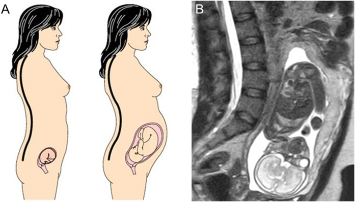
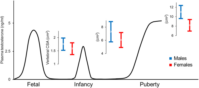
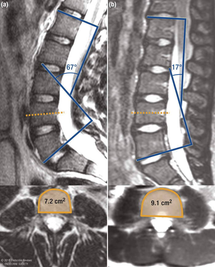
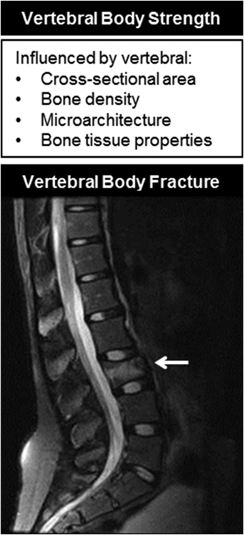
Recent observations indicate that the cross-sectional area (CSA) of vertebral bodies is on average 10% smaller in healthy newborn girls than in newborn boys, a striking difference that increases during infancy and puberty and is greatest by the time of sexual and skeletal maturity. The smaller CSA of female vertebrae is associated with greater spinal flexibility and could represent the human adaptation to fetal load in bipedal posture. Unfortunately, it also imparts a mechanical disadvantage that increases stress within the vertebrae for all physical activities. This review summarizes the potential endocrine, genetic, and environmental determinants of vertebral cross-sectional growth and current knowledge of the association between the small female vertebrae and greater risk for a broad array of spinal conditions across the lifespan.


Figures

Figure 1.
Figure 1.
(a) Schematic representation of increasing lumbar lordosis during pregnancy to compensate for fetal load and maintain a stable position of the center of mass (black and white circle with crosshairs). Adapted from (28). (b) MRI image of a 30-year-old woman at 25 weeks’ gestation showing marked lordosis. Figure 1a adapted with permission from Macmillan Publishers Ltd: Whitcome KA, Shapiro LJ, Lieberman DE. Fetal load and evolution of lumbar lordosis in bipedal hominins. Nature. 2007; 450:7172.
Figure 2.
Figure 2.
Representation of plasma testosterone levels in the male fetus, infant, and adolescent (83), with overlying values for vertebral CSA (mean ± standard deviation) displaying increasing sex differences from birth to prepubertal and postpubertal stages. Female values are ~10% smaller at birth, 15% smaller during childhood, and 25% smaller at sexual maturity and young adulthood (27, 93, 134).
Figure 3.
Figure 3.
Computed tomography measures of vertebral bone density in male (blue) and female (red) subjects, aged 7 to 20 years, showing no sex differences in bone density during the first decade of life and in young adulthood. Although gains in bone density during sexual development are similar for both sexes, the increase begins and reaches peak values at an earlier age in female subjects, consistent with differences in pubertal timing. Data from (147).
Figure 4.
Figure 4.
Longitudinal measurements of vertebral CSA in 20 girls from the commencement of puberty (Tanner stage 2) to its completion (Tanner stage 5). Yellow lines represent changes in values for the children in the lowest baseline quartiles; purple and green lines depict changes in values for the girls in the middle quartiles at baseline; blue lines represent changes in values for the girls in the highest baseline quartiles. Values are shown (a) for each girl and (b) for each quartile. The regression lines across Tanner stages for this phenotype differed between quartiles and did not overlap. Adapted with permission from Loro ML, Sayre J, Roe TF, Goran MI, Kaufman FR, Gilsanz V. Early identification of children predisposed to low peak bone mass and osteoporosis later in life. J Clin Endocrinol Metab. 2000;85:3908–3918.
Figure 5.
Figure 5.
MRI images of the lumbar spine showing a greater LL angle (67° vs 17°) and smaller fourth lumbar vertebral CSA (7.2 cm2 vs 9.1 cm2) in (a) a 12-year-old girl with spondylolysis and (b) a 12-year-old girl without spondylolysis.
Figure 6.
Figure 6.
The strength of the vertebral body is determined by vertebral CSA, bone density, microarchitecture, and bone tissue properties. Fractures (arrow) occur when the loads applied to a vertebral body exceed its strength.

References

    1. Dennison EM, Harvey NC, Cooper C. Programming of osteoporosis and impact on osteoporosis risk. Clin Obstet Gynecol. 2013;56(3):549–555. - PMC - PubMed
    1. Wood CL, Stenson C, Embleton N. The developmental origins of osteoporosis. Curr Genomics. 2015;16(6):411–418. - PMC - PubMed
    1. Antoniades L, MacGregor AJ, Andrew T, Spector TD. Association of birth weight with osteoporosis and osteoarthritis in adult twins. Rheumatology (Oxford). 2003;42(6):791–796. - PubMed
    1. Dennison EM, Syddall HE, Sayer AA, Gilbody HJ, Cooper C. Birth weight and weight at 1 year are independent determinants of bone mass in the seventh decade: the Hertfordshire cohort study. Pediatr Res. 2005;57(4):582–586. - PubMed
    1. Cooper C, Westlake S, Harvey N, Javaid K, Dennison E, Hanson M. Review: developmental origins of osteoporotic fracture. Osteoporos Int. 2006;17(3):337–347. - PubMed

Publication types