

Comparative Genotoxicity of TEMPO and 3 of Its Derivatives in Mouse Lymphoma Cells
- PMID: 29385624
- PMCID: PMC6388407
- DOI: 10.1093/toxsci/kfy022
Comparative Genotoxicity of TEMPO and 3 of Its Derivatives in Mouse Lymphoma Cells
Abstract
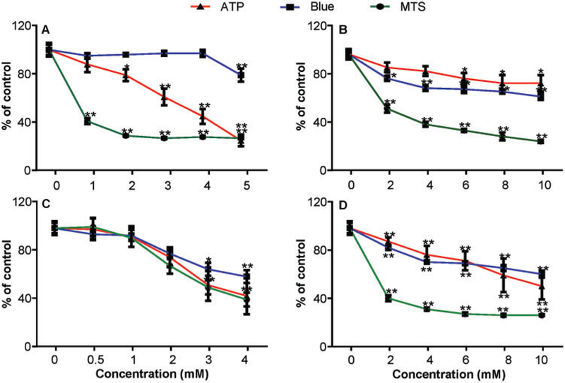
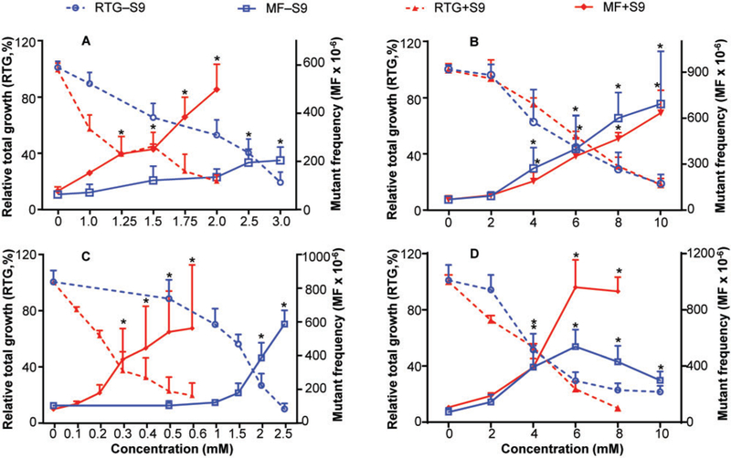
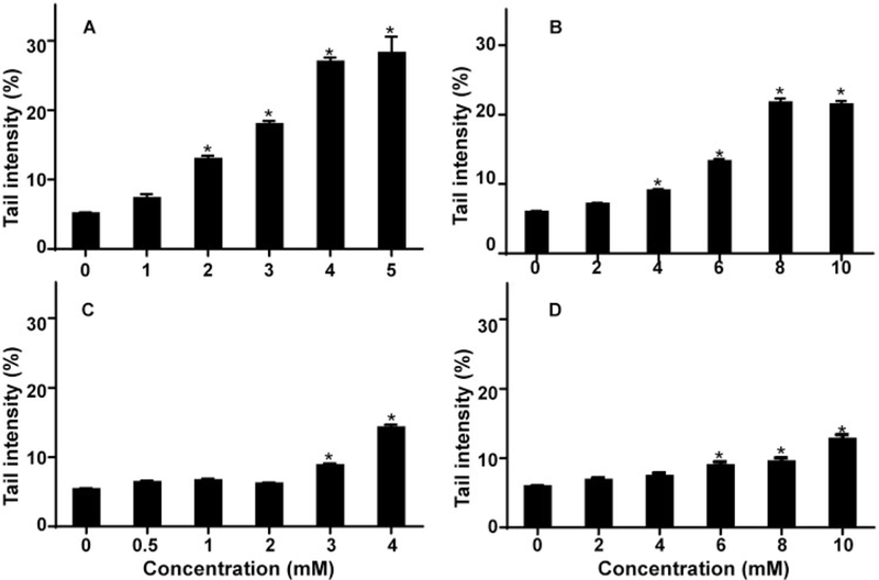
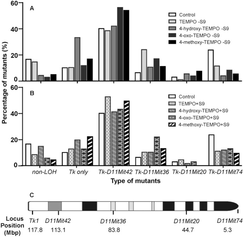
TEMPO (2, 2, 6, 6-tetramethylphiperidine-1-oxyl) and its derivatives are stable free radical nitroxides widely used in the field of chemistry, biology, and pharmacology. TEMPO was previously found to be mutagenic and to induce micronuclei in mammalian cells. In this study, we investigated and quantified the genotoxicity of 4 structurally similar nitroxides, TEMPO and 3 of its derivatives (4-hydroxy-TEMPO, 4-oxo-TEMPO, and 4-methoxy-TEMPO), using the mouse lymphoma assay (MLA) and Comet assay in L5178Y Tk+/- cells. The results showed that all tested nitroxides were cytotoxic and mutagenic in the MLA, both in the presence and absence of S9, with metabolic activation significantly enhancing the cytotoxicity and/or mutagenicity. In addition, the 4 nitroxides caused DNA-strand breakage. The mutagenicity and DNA damaging dose-responses of the test articles were compared using the PROAST benchmark dose software package. The potency ranking of the 4 nitroxides for mutagenicity was different from the ranking of the DNA damaging effects. The mode of action analysis by a multi-endpoint DNA damage pathway assay classified all 4 nitroxides as clastogens. In addition, the majority of the induced Tk mutants showed loss of heterozygosity at the Tk and D11Mit42 loci (ie, chromosome damage <31 Mbp). These results suggest that TEMPO and its 3 derivatives are cytotoxic and mutagenic in mouse lymphoma cells through a mechanism that involves strand breakage and large alterations to DNA. The potency rankings indicate that the different TEMPO derivatives vary in their mutagenic and DNA damaging potential.
Figures
Similar articles
-
Nitroxide TEMPO: a genotoxic and oxidative stress inducer in cultured cells.Toxicol In Vitro. 2013 Aug;27(5):1496-502. doi: 10.1016/j.tiv.2013.02.019. Epub 2013 Mar 18. Toxicol In Vitro. 2013. PMID: 23517621 Free PMC article.
-
Mutant frequency in comparison to oxidative DNA damage induced by ochratoxin A in L5178Y tk+/- (3.7.2C) mouse lymphoma cells.Drug Chem Toxicol. 2014 Apr;37(2):227-32. doi: 10.3109/01480545.2013.838775. Epub 2013 Oct 28. Drug Chem Toxicol. 2014. PMID: 24164384
-
Quantitative analysis of the relative mutagenicity of five chemical constituents of tobacco smoke in the mouse lymphoma assay.Mutagenesis. 2016 May;31(3):287-96. doi: 10.1093/mutage/gev039. Epub 2015 May 22. Mutagenesis. 2016. PMID: 26001754 Free PMC article.
-
Quantitative differentiation of whole smoke solution-induced mutagenicity in the mouse lymphoma assay.Environ Mol Mutagen. 2018 Mar;59(2):103-113. doi: 10.1002/em.22151. Epub 2017 Nov 9. Environ Mol Mutagen. 2018. PMID: 29119619 Free PMC article.
-
Genotoxic and mutagenic potential of camphorquinone in L5178/TK+/- mouse lymphoma cells.Dent Mater. 2018 Mar;34(3):519-530. doi: 10.1016/j.dental.2017.12.013. Epub 2018 Jan 17. Dent Mater. 2018. PMID: 29373133
Cited by
-
Comparative potency analysis of whole smoke solutions in the bacterial reverse mutation test.Mutagenesis. 2021 Aug 27;36(4):321-329. doi: 10.1093/mutage/geab021. Mutagenesis. 2021. PMID: 34131742 Free PMC article.
-
Mechanistic Evaluation of Black Cohosh Extract-Induced Genotoxicity in Human Cells.Toxicol Sci. 2021 Jul 16;182(1):96-106. doi: 10.1093/toxsci/kfab044. Toxicol Sci. 2021. PMID: 33856461 Free PMC article.
-
Spatial-Selective Volumetric 4D Printing and Single-Photon Grafting of Biomolecules within Centimeter-Scale Hydrogels via Tomographic Manufacturing.Adv Mater Technol. 2023 May 23;8(15):admt.202300026. doi: 10.1002/admt.202300026. eCollection 2023 Aug. Adv Mater Technol. 2023. PMID: 37811162 Free PMC article.
-
Effect of Low Concentration of Nitroxides on SH-SY5Y Cells Transfected with the Tau Protein.Int J Mol Sci. 2023 Nov 23;24(23):16675. doi: 10.3390/ijms242316675. Int J Mol Sci. 2023. PMID: 38069000 Free PMC article.
-
Optimized Photoclick (Bio)Resins for Fast Volumetric Bioprinting.Adv Mater. 2021 Dec;33(49):e2102900. doi: 10.1002/adma.202102900. Epub 2021 Oct 5. Adv Mater. 2021. PMID: 34611928 Free PMC article.
References
-
- Adams WT, and Skopek TR (1987). Statistical test for the comparison of samples from mutational spectra. J. Mol. Biol. 194, 391–396. - PubMed
Publication types
MeSH terms
Substances
Associated data
Grants and funding
LinkOut - more resources
Full Text Sources
Other Literature Sources
Miscellaneous
