Review
. 2018 Jan 30;19(2):407.
doi: 10.3390/ijms19020407.

Electrospinning of Chitosan-Based Solutions for Tissue Engineering and Regenerative Medicine

Affiliations
Review

Electrospinning of Chitosan-Based Solutions for Tissue Engineering and Regenerative Medicine

Saad B Qasim et al. Int J Mol Sci. .

Abstract

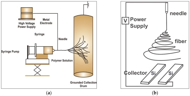
Electrospinning has been used for decades to generate nano-fibres via an electrically charged jet of polymer solution. This process is established on a spinning technique, using electrostatic forces to produce fine fibres from polymer solutions. Amongst, the electrospinning of available biopolymers (silk, cellulose, collagen, gelatine and hyaluronic acid), chitosan (CH) has shown a favourable outcome for tissue regeneration applications. The aim of the current review is to assess the current literature about electrospinning chitosan and its composite formulations for creating fibres in combination with other natural polymers to be employed in tissue engineering. In addition, various polymers blended with chitosan for electrospinning have been discussed in terms of their potential biomedical applications. The review shows that evidence exists in support of the favourable properties and biocompatibility of chitosan electrospun composite biomaterials for a range of applications. However, further research and in vivo studies are required to translate these materials from the laboratory to clinical applications.

Keywords: chitosan; composite solutions; electrospinning; regeneration; tissue engineering.


Conflict of interest statement

The authors declare no conflict of interest.

Figures

Figure 1
Figure 1
The electrospinning process shown schematically (a) electrospinning equipment plate or rotating mandrel (b) aligned collection plates for electrospun nano-fibres [7].
Figure 2
Figure 2
Chemical structure of chitosan showing amide and hydroxyl group that can react and readily form bonds with other natural or synthetic polymers/biomolecules [44].
Figure 3
Figure 3
Illustration depicting the deacetylation process adapted to extract CH from chitin [45].
Figure 4
Figure 4
Chitosan PEO nano-fibres depicting the effects of acetic acid concentration, (a) 2:3 CH:PEO in 45% (b) 4:9 CH:PEO in 36%; (c) 2:3 CH:PEO with 2.5 wt.% total polymer 40%; (d) 4:9 CH:PEO with 2.6 wt.% total polymer 32%; (e) 2:3 CH:PEO with 3 wt.% total polymer blend and 32%; (f) 8:9 CH:PEO with 3.4 wt.% total polymer 32% of total acetic acid concentration [59]; scale bar represents 1 μm (Adapted with permission from publisher).
Figure 5
Figure 5
SEM images of electrospun CH-PEO blends; 4.5 wt.% CH:PEO 95:5, and 10:1, 3 wt.% acetic acid in Dimethyl Sulphoxide (DMSO), chitosan electrospun fibres spun using PEO as co-polymer. (a) Overly aligned fibres (b) random fibres (c,d) fibre distribution frequency calculated from 100 fibres (e,f) orientation histograms showing distribution of aligned and random fibres. Image adapted with permission from publisher (scale bar = 5 µm) [3].
Figure 6
Figure 6
Proposed hydrogen bonding interactions of PEO and CH [125]; (Adapted with permission of publisher).
Figure 7
Figure 7
(A) SEM micrographs of nanohydroxyapatite/collagen/chitosan fibres; scale bar: 1 µm (B) Induced pluripotent stem-cell-derived mesenchymal stem cells (iPSC-MSCs) seeded on HA/chitosan fibres after culturing for 4 days, scale bar: 20 µm (Xie et al., 2016) [142]; (Adapted with permission of publisher).
Figure 8
Figure 8
(A) Macroscopic image of chitosan fibre and (B) fibrous mat; (C) Morphology of fibre evaluated by SEM and atomic force microscopy of 0.1% genipin crosslinked and 1% HA loaded; (D) 7% chitosan fibres, typical morphology seen inset images [143]; (Adapted with permission of publisher).

Similar articles

Cited by

References

    1. Subbiah T., Bhat G.S., Tock R.W., Parameswaran S., Ramkumar S.S. Electrospinning of nanofibers. J. Appl. Polym. Sci. 2005;96:557–569. doi: 10.1002/app.21481. - DOI
    1. Saito M., Tsuji T. Extracellular matrix administration as a potential therapeutic strategy for periodontal ligament regeneration. Expert Opin. Biol. Ther. 2012;12:299–309. doi: 10.1517/14712598.2012.655267. - DOI - PubMed
    1. Qasim S.B., Najeeb S., Delaine-Smith R.M., Rawlinson A., Ur Rehman I. Potential of electrospun chitosan fibers as a surface layer in functionally graded GTR membrane for periodontal regeneration. Dent. Mater. 2016;33:71–83. doi: 10.1016/j.dental.2016.10.003. - DOI - PubMed
    1. Sell S.A., Wolfe P.S., Garg K., McCool J.M., Rodriguez I.A., Bowlin G.L. The use of natural polymers in tissue engineering: A focus on electrospun extracellular matrix analogues. Polymers. 2010;2:522–553. doi: 10.3390/polym2040522. - DOI
    1. Bhardwaj N., Kundu S.C. Electrospinning: A fascinating fiber fabrication technique. Biotechnol. Adv. 2010;28:325–347. doi: 10.1016/j.biotechadv.2010.01.004. - DOI - PubMed

LinkOut - more resources