

Autosomal Recessive NRL Mutations in Patients with Enhanced S-Cone Syndrome
- PMID: 29385733
- PMCID: PMC5852564
- DOI: 10.3390/genes9020068
Autosomal Recessive NRL Mutations in Patients with Enhanced S-Cone Syndrome
Erratum in
-
Correction: Littink, K. W.; et al. Autosomal Recessive NRL Mutations in Patients with Enhanced S-Cone Syndrome. Genes 2018, 9, 68.Genes (Basel). 2018 Mar 7;9(3):145. doi: 10.3390/genes9030145. Genes (Basel). 2018. PMID: 29518905 Free PMC article.
Abstract
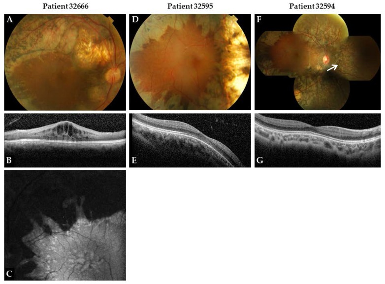
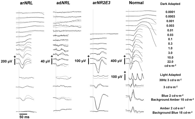
Enhanced S-cone syndrome (ESCS) is mainly associated with mutations in the NR2E3 gene. However, rare mutations in the NRL gene have been reported in patients with ESCS. We report on an ESCS phenotype in additional patients with autosomal recessive NRL (arNRL) mutations. Three Moroccan patients of two different families with arNRL mutations were enrolled in this study. The mutation in the DNA of one patient, from a consanguineous marriage, was detected by homozygosity mapping. The mutation in the DNA of two siblings from a second family was detected in a targeted next-generation sequencing project. Full ophthalmic examination was performed, including best-corrected visual acuity, slit-lamp biomicroscopy, funduscopy, Goldmann kinetic perimetry, optical coherence tomography, fundus autofluorescence, and extended electroretinography including an amber stimulus on a blue background and a blue stimulus on an amber background. One patient carried a homozygous missense mutation (c.508C>A; p.Arg170Ser) in the NRL gene, whereas the same mutation was identified heterozygously in the two siblings of a second family, in combination with a one base-pair deletion (c.654del; p.Cys219Valfs*4) on the other allele. All patients had reduced visual acuity and showed a typical clumped pigmentary retinal degeneration (CPRD). Foveal schisis-like changes were observed in the oldest patient. An electroretinogram (ERG) under dark-adapted conditions showed absent responses for low stimulus strengths and reduced responses for high stimulus strengths, with constant b-wave latencies despite increasing stimulus strength. A relatively high amplitude was detected with a blue stimulus on an amber background, while an amber stimulus on a blue background showed reduced responses. The arNRL mutations cause a phenotype with typical CPRD. This phenotype has previously been described in patients with ESCS caused by NR2E3 mutations, and rarely by NRL mutations. Based on our findings in ERG testing, we conclude that S-cone function is enhanced in our patients in a similar manner as in patients with NR2E3-associated ESCS, confirming previous reports of NRL as a second gene to cause ESCS.
Keywords: S-cone-specific ERG; autosomal recessive NRL; enhanced S-cone syndrome; hereditary retinal dystrophy.
Conflict of interest statement
The authors declare no conflict of interest. The founding sponsors had no role in the design of the study; in the collection, analyses, or interpretation of data; in the writing of the manuscript, and in the decision to publish the results.
Figures
References
-
- Milam A.H., Rose L., Cideciyan A.V., Barakat M.R., Tang W.X., Gupta N., Aleman T.S., Wright A.F., Stone E.M., Sheffield V.C., et al. The nuclear receptor NR2E3 plays a role in human retinal photoreceptor differentiation and degeneration. Proc. Natl. Acad. Sci. USA. 2002;99:473–478. doi: 10.1073/pnas.022533099. - DOI - PMC - PubMed
-
- Jacobson S.G., Marmor M.F., Kemp C.M., Knighton R.W. SWS (blue) cone hypersensitivity in a newly identified retinal degeneration. Investig. Ophthalmol. Vis. Sci. 1990;31:827–838. - PubMed
LinkOut - more resources
Full Text Sources
Other Literature Sources
