. 2018 Feb 28;10(8):6904-6916.
doi: 10.1021/acsami.7b18525. Epub 2018 Feb 13.

Nitric Oxide-Delivering High-Density Lipoprotein-like Nanoparticles as a Biomimetic Nanotherapy for Vascular Diseases

Affiliations

Nitric Oxide-Delivering High-Density Lipoprotein-like Nanoparticles as a Biomimetic Nanotherapy for Vascular Diseases

Jonathan S Rink et al. ACS Appl Mater Interfaces. .

Abstract

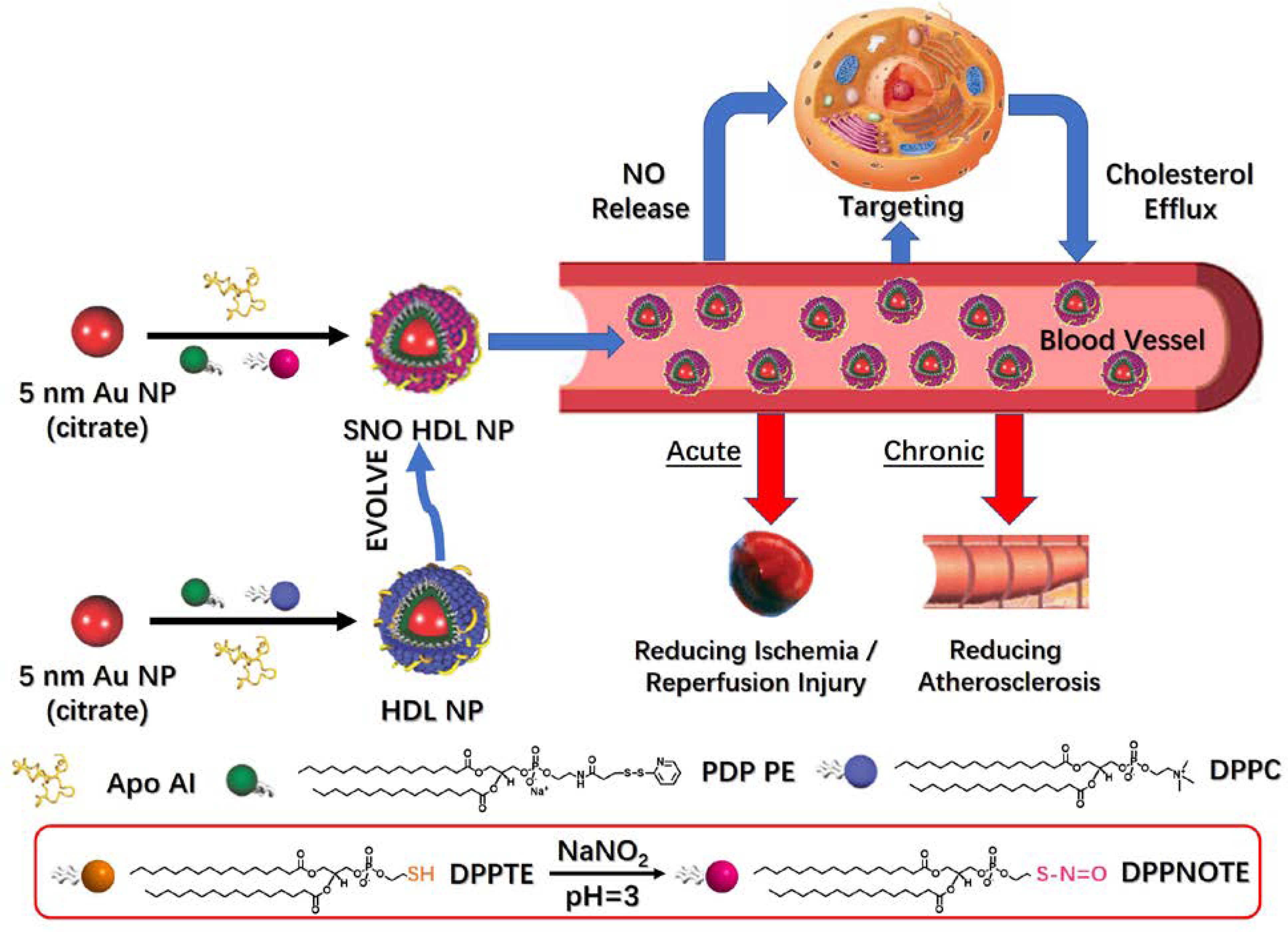
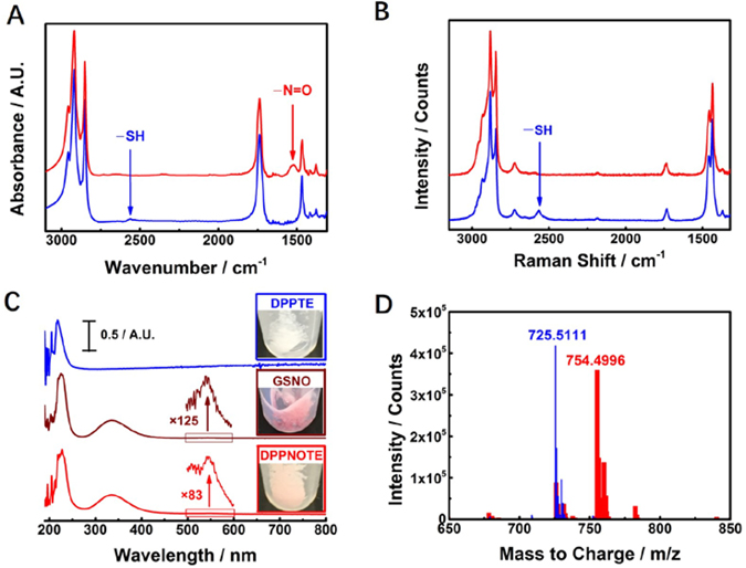
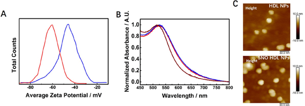
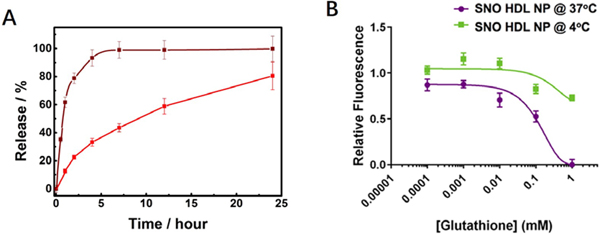
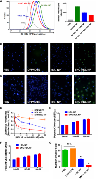
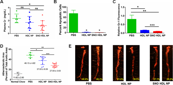
Disorders of blood vessels cause a range of severe health problems. As a powerful vasodilator and cellular second messenger, nitric oxide (NO) is known to have beneficial vascular functions. However, NO typically has a short half-life and is not specifically targeted. On the other hand, high-density lipoproteins (HDLs) are targeted natural nanoparticles (NPs) that transport cholesterol in the systemic circulation and whose protective effects in vascular homeostasis overlap with those of NO. Evolving the AuNP-templated HDL-like nanoparticles (HDL NPs), a platform of bioinspired HDL, we set up a targeted biomimetic nanotherapy for vascular disease that combines the functions of NO and HDL. A synthetic S-nitrosylated (SNO) phospholipid (1,2-dipalmitoyl-sn-glycero-3-phosphonitrosothioethanol) was synthesized and assembled with S-containing phospholipids and the principal protein of HDL, apolipoprotein A-I, to construct NO-delivering HDL-like particles (SNO HDL NPs). SNO HDL NPs self-assemble under mild conditions similar to natural processes, avoiding the complex postassembly modification needed for most synthetic NO-release nanoparticles. In vitro data demonstrate that the SNO HDL NPs merge the functional properties of NO and HDL into a targeted nanocarrier. Also, SNO HDL NPs were demonstrated to reduce ischemia/reperfusion injury in vivo in a mouse kidney transplant model and atherosclerotic plaque burden in a mouse model of atherosclerosis. Thus, the synthesis of SNO HDL NPs provides not only a bioinspired nanotherapy for vascular disease but also a foundation to construct diversified multifunctional platforms based on HDL NPs in the future.

Keywords: S-nitrosylation; biomimetic; high-density lipoprotein-like nanoparticles; nanotherapy; nitric oxide-delivering; vascular disease.


Figures

Figure 1.
Figure 1.
Preparation, properties and functions of SNO HDL NPs.
Figure 2.
Figure 2.
Characterization of DPPNOTE. A) FTIR spectra of DPPTE (blue solid-line) and DPPNOTE (red solid-line). B) Raman spectra of DPPTE (blue solid-line) and DPPNOTE (red solid-line). C) Normalized UV-Vis spectra of DPPTE (blue solid-line with an inlet of a picture of DPPTE powder), GSNO [brown solid-line with inlets of a magnified spectrum (by 125 folds) ranged from 500 ~ 600 nm and a picture of GSNO powder] and DPPNOTE [red solid-line with inlets of a magnified spectrum (by 83 folds) ranged from 500 ~ 600 nm and a picture of DPPNOTE powder]. D) Mass spectra of DPPTE (narrow blue bars, the dominant peak ascribe to the molecular ion, [DPPTE + NH3]+) and DPPNOTE (broad red bars, the dominant peak ascribe to the molecular ion, [DPPNOTE + NH3]+).
Figure 3.
Figure 3.
Characterization of SNO HDL NPs. A) Zeta potentials of HDL NPs (blue solid-line) and SNO HDL NPs (red solid-line). B) Normalized UV-Vis spectra of bare 5 nm AuNPs (brown solid-line), HDL NPs (blue solid-line) and SNO HDL NPs DPPTE (red solid-line). C) AFM images of HDL NPs and SNO HDL NPs.
Figure 4.
Figure 4.
In vitro characterization of NO Release from SNO HDL NPs. A) NO release from SNO HDL NPs (red solid-line) and DPPNOTE (brown solid-line) at 37oC. B) GSH -induced NO release from SNO HDL NPs at 4 °C and 37 °C.
Figure 5.
Figure 5.
In vitro characterization of SNO HDL NP delivery of NO and function. A) Competitive binding of SNO HDL NPs and HDL NPs to macrophage cells. Left- representative histogram of DiI HDL NP fluorescence of THP-1 cells treated with DiI HDL NPs alone, or in combination with unlabeled HDL NPs or SNO HDL NPs. Right- median fluorescent intensities for each treatment group. B, C) Release of NO to J774 macrophages (B) and differentiated THP-1 macrophages (C) treated with DAF FM diacetate. From left to right: PBS, DPPNOTE (2 μM), HDL NP (25 nM), and SNO HDL NP (25 nM). Green- DAF FM diacetate. Blue- nuclei (DAPI). Images taken at 40X magnification. D) Quantiblue assay of THP-1 Dual cells treated with 5ng/ mL LPS and increasing concentrations of DPPNOTE, HDL NP or SNO HDL NPs. *p<0.05 vs. all other treatment groups. E, F) Cholesterol efflux from 3H-cholesterol loaded macrophages to SNO HDL NPs. J774 macrophages (E) and differentiated THP-1 macrophages (F) were loaded with 3H-cholesterol prior to addition of cholesterol acceptors for 24 hrs. G) SNO HDL NPs reduced transwell migration of AoSMCs compared with controls, PBS, DPPNOTE alone and HDL NPs (*p<0.0001 v. all other treatment groups).
Figure 6.
Figure 6.
SNO HDL NPs as therapy for IRI and atherosclerosis. A) Plasma creatinine levels of mouse kidney transplant recipients on day 2 post transplantation (*p=0.0113 v. PBS control). B) Quantification of apoptosis (TUNEL) staining in kidney transplant recipients (*p<0.0001 vs. PBS). C) Quantification of immunocytochemistry staining for Gr-1, a neutrophil marker, in kidney transplant recipients (*p=0.0175. **p=0.0041. ***p=0.008). D) ApoE knockout mice were fed a high fat diet for 12 weeks, then administered PBS, HDL NPs or SNO HDL NPs 3×/ week for an additional 6 weeks. Atherosclerotic lesions were visualized using Sudan Red staining. The atherosclerotic area was calculated by measuring the area of Sudan Red staining, divided by the total aortic area (*p=0.0149. **p<0.0001. ***p=0.0118). E) Representative images of aortas from mice treated with PBS, HDL NP or SNO HDL NPs. Red fluorescent staining indicates atherosclerotic plaques. Images taken at 6X magnification.

References

    1. Roger VL; Go AS; Lloyd-Jones DM; Adams RJ; Berry JD; Brown TM; Camethon MR; Dai S; de Simone G; Ford ES; Fox CS; Fullerton HJ; Gillespie C; Greenlund KJ; Hailpem SM; Heit JA; Ho PM; Howard VJ; Kissela BM; Kittner SJ; Lackland DT; Lichtman JH; Lisabeth LD; Makuc DM; Marcus GM; Marelli A; Matchar DB; McDermott MM; Meigs JB; Moy CS; Mozaffarian D; Mussolino ME; Nichol G; Paynter NP; Rosamond WD; Sorlie PD; Stafford RS; Turan TN; Turner MB; Wong ND; Wylie-Rosett J; American Heart Assoc Stat, C.; Stroke Stat, S., Heart Disease and Stroke Statistics-2011 Update A Report From the American Heart Association. Circulation 2011, 123 (4), E18–E209. - PMC - PubMed
    1. Schulz R; Kelm M; Heusch G, Nitric oxide in myocardial ischemia/reperfusion injury. Cardiovascular research 2004, 61 (3), 402–413. - PubMed
    1. Giraldez RR; Panda A; Xia Y; Sanders SP; Zweier JL, Decreased nitric-oxide synthase activity causes impaired endothelium-dependent relaxation in the postischemic heart. Journal of Biological Chemistry 1997, 272 (34), 21420–21426. - PubMed
    1. Napoli C; de Nigris F; Williams-Ignarro S; Pignalosa O; Sica V; Ignarro LJ, Nitric oxide and atherosclerosis: an update. Nitric oxide 2006, 15 (4), 265–279. - PubMed
    1. Garg UC; Hassid A, Nitric oxide-generating vasodilators and 8-bromo-cyclic guanosine monophosphate inhibit mitogenesis and proliferation of cultured rat vascular smooth muscle cells. Journal of Clinical Investigation 1989, 83 (5), 1774–1777. - PMC - PubMed