. 2018 Jun;33(4):221-231.
doi: 10.1177/1533317518755993. Epub 2018 Jan 31.

The Role of Musk in Relieving the Neurodegenerative Changes Induced After Exposure to Chronic Stress

Affiliations

The Role of Musk in Relieving the Neurodegenerative Changes Induced After Exposure to Chronic Stress

Manal Galal Abd El Wahab et al. Am J Alzheimers Dis Other Demen. 2018 Jun.

Abstract

Objective: This study aimed to evaluate the effect induced by musk on Alzheimer's disease-such as neurodegenerative changes in mice exposed to chronic unpredictable mild stress (CUMS).

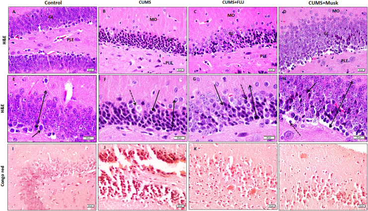
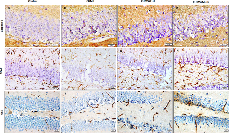
Material and methods: Forty male Swiss albino mice were divided into 4 groups (n = 10); control, CUMS, CUMS + fluoxetine, CUMS + musk. At the end of the experiment, behavior of the mice was assessed. Serum corticosterone level, hippocampal protein level of the glucocorticoid receptors, and brain-derived neurotropic factor were also assessed. Hippocampus was histopathologically examined.

Results: Musk improved depressive status induced after exposure to CUMS as evidenced by the forced swimming and open field tests and improved the short-term memory as evidenced by the elevated plus maze test. Musk reduced both corticosterone levels and the hippocampal neurodegenerative changes observed after exposure to CUMS. These improvements were comparable to those induced by fluoxetine.

Conclusion: Musk alleviated the memory impairment and neurodegenerative changes induced after exposure to the chronic stress.

Keywords: Alzheimer; GR-BDNF; chronic stress; corticosterone; depression; musk; neurodegenerative.


Conflict of interest statement

The authors declared no potential conflicts of interest with respect to the research, authorship, and/or publication of this article.

Figures

Figure 1.
Figure 1.
Procedure of the experiment. Mice were exposed to chronic unpredictable mild stress (CUMS) from day 1 to day 28. On day 29 to day 42, the mice were exposed daily to the control treatment (amyl acetate), fluoxetine, or muck. On the days 43 to 45, the behavioral tests were sequentially done for all groups. On the day 46, blood samples were obtained then the mice were sacrificed.
Figure 2.
Figure 2.
Forced swimming test (A) and open field test (B, C) of the control, chronic unpredictable mild stress (CUMS), fluoxetine-treated (CUMS + FLU), and musk-treated (CUMS + M) groups (n = 10 each). Data were shown as mean (SD). # indicates significance compared to the control group; *indicates significance compared to the CUMS group.
Figure 3.
Figure 3.
A, Serum level of corticosterone shown as mean (SD). Glucocorticoid receptor protein (B) and BDNF protein (C) expression levels in the hippocampus assessed by ELISA and expressed as percentage of control value (SD; n = 10 each). #indicates significance compared to the control group; *indicates significance compared to the CUMS group. BDNF indicates brain-derived neurotropic factor; CUMS, chronic unpredictable mild stress, ELISA, enzyme-linked immunosorbent assay; FLU, fluoxetine; M, musk; SD, standard deviation.
Figure 4.
Figure 4.
The hippocampal CA1 is formed of the molecular layer (MO), the pyramidal layer (PY), and the polymorphic layer (PO). The thickness of the PY layer appears smaller in the chronic unpredictable mild stress (CUMS) compared to the control (bi-head arrow). Some darkly stained cells (interrupted arrow) are observed. An eosinophilic structure (black arrow) is seen related to the pyramidal cell. Amyloid deposition with salmon red coloration observed near CA1 (insert show blood vessel with amyloid deposition in its wall). Immunoexpression of Caspase, GFAP, in the hippocampal CA1 are shown (A-D, I-T x 400, E-Hx1000). CUMS indicates chronic unpredictable mild stress, FLU, fluoxetine; M, musk.
Figure 5.
Figure 5.
Thickness and surface area of CA1 (A) and dentate gyrus (B). Immunoexpression of GFAP (C), Caspase-3 (D), Ki67 (E), in the studied groups. Data are expressed as mean. # indicates significance versus control; * indicates significance versus CUMS. CA indicates cornu ammonis; CUMS, chronic unpredictable mild stress; FLU, fluoxetine; M, musk.
Figure 6.
Figure 6.
The hippocampal DG is formed of the PLE, the GL, and the MO. The thickness of the GL layer appears smaller in the CUMS compared to the control (bi-head arrow). Some cells appear vacuolated cells (interrupted arrow) are observed. Note the increased number of the immature cell that have darkly stained nuclei (arrow). Amyloid deposition with salmon red coloration observed as patches as well as aggregation around the granular cell of the DG (A-D, I-L x400, E-H x1000). CUMS indicates chronic unpredictable mild stress, DG, dentate gyrus; FLU, fluoxetine; GL, granular cell layer; M; musk; MO, molecular; PLE, pleomorphic layer.
Figure 7.
Figure 7.
Immunoexpression of GFAP, Caspase-3, Ki67, in the hippocampal dentate gyrus (A-D x600 and E-L x400). CUMS indicates chronic unpredictable mild stress; FLU, fluoxetine; M, musk.

References

    1. World Alzheimer Report 2016. Improving healthcare for people living with dementia coverage, Quality and costs now and in the future. Alzheimer’s Disease International (ADI), London. https://www.alz.co.uk/research/WorldAlzheimerReport2016.pdf Accessed January 19, 2018.
    1. World Alzheimer Report 2015. The Global Impact of Dementia An analysis of revalence, incidence, cost and trends. Executive summary. Alzheimer’s Disease International (ADI), London. https://www.alz.co.uk/research/worldalzheimerreport2015summary.pdf Accessed January 19, 2018.
    1. Yamada T, Hattori H, Miura A, Tanabe M, Yamori Y. Prevalence of Alzheimer’s disease, vascular dementia and dementia with Lewy bodies in a Japanese population. Psychiatry Clin Neurosci. 2001;55(1):21–25. - PubMed
    1. Chung JK, Plitman E, Nakajima S, et al. ; Alzheimer’s Disease Neuroimaging Initiative. Cortical amyloid β deposition and current depressive symptoms in Alzheimer disease and mild cognitive impairment. J Geriatr Psychiatry Neurol. 2016;29(3):149–159. - PMC - PubMed
    1. Choudhury B, Saytode P, Shah V. Neurodegenrative disorders: past, present and future. Int J Appl Biol Pharma Technol. 2014;5(2):1–14.

Publication types

MeSH terms