

Magnetic resonance-guided interstitial high-intensity focused ultrasound for brain tumor ablation
- PMID: 29385926
- PMCID: PMC5907801
- DOI: 10.3171/2017.11.FOCUS17613
Magnetic resonance-guided interstitial high-intensity focused ultrasound for brain tumor ablation
Abstract
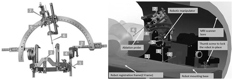
Currently, treatment of brain tumors is limited to resection, chemotherapy, and radiotherapy. Thermal ablation has been recently explored. High-intensity focused ultrasound (HIFU) is being explored as an alternative. Specifically, the authors propose delivering HIFU internally to the tumor with an MRI-guided robotic assistant (MRgRA). The advantage of the authors' interstitial device over external MRI-guided HIFU (MRgHIFU) is that it allows for conformal, precise ablation and concurrent tissue sampling. The authors describe their workflow for MRgRA HIFU delivery.
Keywords: DOF = degrees of freedom; GBM = glioblastoma; HIFU = high-intensity focused ultrasound; LITT = laser interstitial thermal therapy; MRI guided; MRIT = MRI thermometry; MRgHIFU = MRI-guided HIFU; MRgRA = MRI-guided robot assistant; SRS = stereotactic radiosurgery; brain tumor; high-intensity focused ultrasound; neural ablation.
Figures

References
-
- Canney MS, Chavrier F, Tsysar S, Chapelon JY, Lafon C, Carpentier A. A multi-element interstitial ultrasound applicator for the thermal therapy of brain tumors. J Acoust Soc Am. 2013;134:1647–1655. - PubMed
-
- Chen X, Diederich CJ, Wootton JH, Pouliot J, Hsu IC. Optimisation-based thermal treatment planning for catheter-based ultrasound hyperthermia. Int J Hyperthermia. 2010;26:39–55. - PubMed
-
- Christian E, Yu C, Apuzzo MLJ. Focused ultrasound: relevant history and prospects for the addition of mechanical energy to the neurosurgical armamentarium. World Neurosurg. 2014;82:354–365. - PubMed
-
- Cohen-Inbar O, Melmer P, Lee CC, Xu Z, Schlesinger D, Sheehan JP. Leukoencephalopathy in long term brain metastases survivors treated with radiosurgery. J Neurooncol. 2016;126:289–298. - PubMed
Publication types
MeSH terms
Grants and funding
LinkOut - more resources
Full Text Sources
Other Literature Sources
Medical
